Raising The Bar For Breast Imaging, with Louise Miller of Mammography Educators
Episode Overview
Episode Topic: In this episode of Skeleton Crew – The Rad Tech Show, we delve into the role of Louise Miller, co-founder and director of education at Mammography Educators, who provides a comprehensive overview of the evolution of mammography education and the advancements in breast imaging. The discussion covers the early days of mammography, the initiation of the first mammography class, and the crucial role of the Mammography Quality Standards Act (mQSA) in shaping education. The focus is on the unique approach to education, personal and emotional aspects of mammography, and the data-driven positioning technique known as the “Miller method.”
Lessons You’ll Learn: In this episode, we gain valuable insights into the challenges faced by early mammographers, the development of educational standards, and the global landscape of mammography. The episode highlights the significance of a comprehensive and empathetic approach to education, the role of advanced technologies like Digital Breast Tomosynthesis (DBT), and the increasing trend of technologists seeking training in breast ultrasound. Additionally, it explores the structure of Mammography Initial Training (MIT) programs and the commitment to continuous improvement.
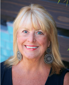
About Our Guests: Join us as we host Louise Miller, with over 25 years of experience, is a co-founder and director of education at Mammography Educators. As a pioneer in mammography education, she has played a vital role in shaping curricula, advocating for breast cancer awareness, and developing positioning techniques. As a breast cancer survivor, Louise brings a unique perspective to the conversation, emphasizing the importance of early detection through mammography.
Topics Covered: The conversation covers a range of topics, including the initiation of the first mammography class, the impact of mQSA on education, the emotional aspects of mammography, and the data-driven “Miller method.” It also delves into advancements like Digital Breast Tomosynthesis, global discrepancies in breast imaging, the role of ultrasound, and the structure of Mammography Initial Training programs. The episode concludes with discussions on personal experiences, ergonomics in mammography, the global shortage of technologists, and practical advice for those entering the field.
Our Guest: Louise Miller, Pioneer in Mammography Education
Louise Miller is a prominent figure in mammography education, boasting more than a quarter-century of impactful teaching. As the co-founder of the Mammography Practicum at UC San Diego, she’s reshaped radiologic technology education, lending her competence to esteemed institutions like Harvard Medical School. Louise’s influence isn’t confined to the US, she’s volunteered in places like Ukraine, contributing significantly to global mammography screening efforts. Her training programmes like the Mammography Initial Training, the Digital Breast Tomosynthesis (DBT) Initial Training, the Breast Ultrasound (Sonography) Initial Training and more, provide flexibility and expert guidance at one’s own convenience, bypassing the constraints of a live course.
Alongside her academic valour, Louise promotes breast care awareness and support through active roles in professional groups such as the American College of Radiology. For her contribution, she received prestigious awards like the NCBC Impact Award in 2011. Despite facing personal health problems, notably Triple Negative Breast Cancer, Louise’s determination to promote breast health remains undeterred.
When not immersed in her professional commitments, Louise finds solace and joy in San Diego’s vibrant community. She’s a passionate mentor, guiding the next generation, and cherishes moments with her supportive family. Her daughter Erin and grandsons Alex and Jeffrey stand by her side, embodying the resilience and dedication that defines Louise’s remarkable journey in mammography education and breast health.


Follow Mammography educators on
Episode Transcript
Louise Miller: Our positioning technique is the only one that’s been proven by data. We actually did a study that was published in the American Journal of Roentgenology in 2017 that showed that our positioning technique improves image quality by up to 50%. Nobody else has that data.
Jennifer Callahan: Welcome to the Skeleton Crew. I’m your host, Jen Callahan, a technologist with ten plus years experience. In each episode, we will explore the fast-paced, ever-changing suburbs, completely crazy field of radiology. We will speak to technologists from all different modalities about their careers and education. The educators and leaders who are shaping the field today, and the business executives whose innovations are paving the future of radiology. This episode is brought to you by X-raytechnicianschools.com. If you’re considering a career in X-ray, visit x-raytechnicianschools.com to explore schools and to get honest information on career paths, salaries, and degree options.
Hey, everybody, welcome back to the Skeleton Crew. I’m your host, Jen Callahan. We’re here to have a great conversation with Louise Miller. We’re going to be talking about the world of mammography. And a little quick background to who Louise is. She is the co-founder and director of education of the Mammography Educators. So thanks, Louise, so much for being with us today.
Louise Miller: Well, thanks, Jen, for having me. What a cool concept this is. I wish this was available when I was growing up as a new radiographer, you know, but nothing like this was available. So this is super cool. Thanks for asking me.
Jennifer Callahan: Oh our pleasure. So, like you were saying, stuff like this wasn’t available, you know, back when you became a radiographer, about how long ago?
Louise Miller: I finished with my RT program in the late 70s. So back in the olden days, before most of you were born or even, you know, thought about going into imaging. Anyway, I started when I started out, I went through the basic program and mammography just kind of got started in the early 80s, and they delivered this machine to our department. And of course, they’re like all the female techs, you’re going to do mammograms and we’re going doing what? We have no idea what it was. And we had an application specialist come in and she showed us basically how to use the machine, but we had no idea what our images were supposed to look like. We had nothing, no background in positioning. We were basically just picking up the breast and throwing it on there and compressing it, hoping for the best. There was no training whatsoever. And actually it was really unfortunate because all the techs hated doing it because we didn’t feel comfortable, we didn’t know how, we didn’t feel confident, and the patients didn’t weren’t very happy. Also because this is when mammography was brand new, so they weren’t used to the idea of having their breasts touched, you know, and positioned by a stranger. And then also the compression thing. So it was like a no-win situation for the patients and the techs. Actually, we all hated doing mammograms so much that we used to say, you really drew the short straw. If you had Barry Minimus in the morning and mammograms in the afternoon. We used to call it butts and boobs and it’s like nobody wanted.
Louise Miller: But what happened is, because there was no training, I got this bright idea to go to the director of the Red tech program at our community college. And I went up there and say, you need to offer a class in mammography. And she goes, great idea. And she goes, well, who’s going to teach it? And I said, well, a radiologist, of course. And she said, well, why don’t you teach it? And I was like, I don’t have a teacher’s credential. I don’t know how to teach. I’ve never given a lecture in my life. She goes, well, you can do it. We can get you on faculty based on your experience. And I was like, are you kidding? And so I was like, okay, I’ll give it a shot. So we started the first mammography class. Actually, it was a one-credit class, 16 hours connected with an RT program in the United States, and actually probably the world. It was the very first one, and this was in the early 90s. So something I’m super proud of. And now here we are, what, you know, 30 years later and they’re still running that program at the community college. I’ve now passed it on, passed the baton to new, younger technologists who are out teaching. And same with me. You don’t necessarily have to have a teaching credential to create this type of opportunity for yourself, and also make it available to techs who really need it. So that was a real starting point in my career.
Jennifer Callahan: How did you even develop a program or like the credits that you’re going to be teaching? I mean, like you said, you were new to it yourself. You’re looking for guidance from an actual school, and they’re like, we think that you should do this. Where did you gather the information from?
Louise Miller: Basically, I’ve always believed that teaching breast imaging and mammography should be based on the principles of general radiology. You have to know the correlational anatomy and physiology. You have to know the physics involved. You have to understand all the characteristics of everything we do with general Rad applied to mammography. So I kind of use that as my guideline. And still to this day, this is how we’re teaching. We just don’t teach how to do something. We want to understand why they’re doing something before they learn the how. And that’s something that’s very unique to our mammography training program. We’re the only company that does that. We just don’t follow the basic curriculum that’s spelled out in the content specifications by ART or mQSA. We expand it. We want our students to become good mammographer not just pass the test. But another thing that really helped me is that there was some talk about legislation which turned out to be the Mammography Quality Standards Act, which was passed in 1993. So I happened to. By circumstance, odd circumstances. I was a volunteer for the American Cancer Society, and I met some really wonderful people who were interested in developing a curriculum for technologists. So somehow I got in this very beginning committee, and I was the only person who had developed this little curriculum. So that combined with the curriculum we were interested in developing for the American College of Radiology, which followed the mQSA guidelines, it all kind of fit together. It was just a wonderful opportunity to be able to share what we had developed and again, based on general radiology principles, developing a curriculum that made sense to techs.
Jennifer Callahan: Did you suggest then to the people that you were working with like, hey, you should come and check out this course since everyone was hated to do the butts and boobs day.
Louise Miller: Yeah. Well, exactly. Well, what happened is so many texts were so hungry for information. I mean, we filled the class at the community college. It was just full and so we saw the need. Other programs started looking at our program to kind of replicate across the country. So that’s how we got started there. But then when mQSA was passed, they required a 40 hour base education course and ours was only like 16 hours. So I marched myself up to the University of California to one of the directors of radiology. She was a pioneer in breast imaging and mammography in San Diego. And I went up to her name is Dr. Linda Olsen. Wonderful person. And I said, somebody needs to be doing this 40 hour class. Somebody needs to be running a class like this on a university based level. Once again, she goes, well, you can do it. And I was like, no, I’m not on faculty. And she goes, I’ll put you on faculty. I think the moral of the story is, if you see opportunity and need, just go for it. And I’ve had a wonderful career because I just kind of got set up, saw the need and went for it.
Jennifer Callahan: All right. So you had mentioned the mQSA multiple different times. And myself, as someone who has been researching into, you know, starting into mammography, you always see that on websites and you hear, you know, other mammography talk about it. I feel like it’s a more prevalent thing that you’re hearing about. Could you give some information on one? What is that acronym? And two, how was it developed?
Louise Miller: Okay. It stands for the Mammography Quality Standards Act. Actually, it’s really interesting because it’s one of the few, I think, maybe even the only medical image procedure that has government mandates to it. So it’s not, you know, it’s not like CT, MRI, anything like that. It’s unique in that we have federal legislation, actually, there’s other legislation regarding breast cancer detection like the breast density law. But concerning the mQSA, there was a lot of women’s advocacy groups that were really concerned about the low quality of mammography. We were performing mammograms throughout the 80s and into the 90s, but a lot of cancer was missed due to poor quality. Poor quality could be a technical factor or could be positioning. So it was actually under the directive of some people may remember the Susan G. Komen Foundation. It was Susan Komen sister and a couple other very strong breast cancer advocates that petitioned the government to create a law that. Oversaw the quality of mammograms. So that’s how it was created. And they developed a committee. And actually some of my close colleagues were on that initial committee that set out the standards. And one of the standards was the basic 40 hours education. And they developed, again, the curriculum that should cover anatomy, physiology, physics, positioning, you know, breast cancer treatment, all those types of things. So that’s where that was born. And there’s been a couple updates to it along the way, which is good. Every time it’s updated it’s better for the patient. So it’s just assuring quality. That’s the name Mammography Quality Standards Act.
Jennifer Callahan: That’s great. So much work goes into making sure that qualities are kept. And especially too, because how prevalent that breast cancer has become so really reassuring for for a patient who’s going in and having a study done. Yeah.
Louise Miller: In conjunction with that, I should mention the American College of Radiology instituted its accreditation program. So that’s where they kind of manage the quality for mQSA. They work hand in hand. So what we have to do every three years, we have to submit images. We have to make sure all our credentials are updated. And also to we have mQSA inspections. So everything works together to assure that the patient is getting the highest quality of mammogram. Right.
Jennifer Callahan: So taking that the mQSA that obviously goes into how a curriculum is developed for, you know, education for mammography, is it difficult to incorporate that in or I guess maybe that’s really the standard of guidelines of what you have to follow.
Louise Miller: It is. And it is actually they work in conjunction. Again, everything kind of dominoes onto each other. The a part where we take our advanced certification and mammography, okay. You can take it one year after you receive your basic general radiology license. The are developed their own content specifications based on recommendations. So they have again all the criteria and subcategories that are all listed on their website. What we chose to do for our program, which is unique, we not only covered the topics that they need to be familiar with to quote, pass the test. We feel that our program is unique and that we spend a lot of time preparing the student to be a good Mammographer. Passing the test is not the same as being a good mammographer, so a lot of other programs, maybe they’ll do a 1 or 2 hour lecture on positioning and image quality. We have like eight hours of our program now. We cover all the other topics. It’s not like we leave anything out, but we put a lot of emphasis on positioning and image quality, and that’s unique to us. And also we we have topics like patient relations, us developing good communication skills, understanding patient anxiety, which I think a lot of times techs aren’t prepared for. You know, it’s not like doing a hand or a wrist or a foot. It’s a very personal exam, obviously. So there’s a lot of factors that go along with that.
Jennifer Callahan: Yeah, definitely a very personal part of your body. It’s not like you said, like your hand or wrist and everybody saying all the time. Some women are not to say ashamed with their breasts, but, you know, maybe they wish they were bigger. Maybe they wish that they were smaller. Maybe they feel like they have ugly breasts because a woman or a man, you know, you critique your own body. So that’s absolutely part of it. Or maybe they’re coming in and they’ve already had a scan and they’re coming back for additional imaging. So like you said, to touch on the anxiety portion of it.
Louise Miller: Absolutely. And we teach them not only the source of anxiety, like you said, it could be a previous positive finding on their mammogram. They could be a breast cancer survivor, which always heightens a patient’s anxiety. Those body issues being body conscious and self conscious about our bodies is so prevalent in our society, especially when we have Cosmo and, you know, all these girls on the commercial that have these perfect bodies. A lot of women come in and they feel they kind of don’t measure up or compare. So they’re very inhibited. You know, they don’t they don’t want to expose their breasts. And so those are all things we discuss, which is very unique to our program.
Jennifer Callahan: I’m assuming that one, you address positioning within the training course because that’s numero uno. Pretty much you know, x-ray scan memo is learning how to position. But like x-ray I’m assuming that there’s standardized positioning for mammo and for exams and possibly for I guess maybe depending on exactly what you might be looking for, maybe a patient’s coming back for a follow up to a previous scan.
Louise Miller: Right. Jen, It’s so interesting that you bring that up because believe it or not, there’s no standardization.
Jennifer Callahan: Really. Wow.
Louise Miller: No, its basically a free for all you can get in there and position however you want to, because there hasn’t been a lot of leadership from our professional organizations like the American College of Radiology. I think they did a positioning video like 15 years ago. There’s not a lot of standardization. So that’s something I’ve really pushed. And how I’ve tried to make an inroad about this is, is by working on a lot of national committees, like for the Society of Breast Imaging and with the American College of Radiology and with the SRT, and also going out and speaking at national conferences. Anybody asked me to speak, I’m there. I’m happy to talk about it because I know it works. Our positioning technique is unique to us. Actually. It’s kind of funny because my business partner and all my colleagues decided they wanted to call it the Miller method, and actually it is something I developed. It’s not only the positioning technique, but it’s the way I teach positioning. Like I said, it’s all based on correlative anatomy, but the positioning technique is in unique in that it’s extremely proficient. So that means we can get the most breast tissue the first time. It’s efficient. So you can do it faster. We don’t tell our supervisors that you can you can get through it more quickly. And it’s ergonomically sound. And that’s a huge deal in mammography because a lot of texts have developed wrist and shoulder problems.
Louise Miller: It’s a huge problem. It almost as big as a problem with ergonomics and workplace injuries as ultrasound. We just don’t have the data yet. So our technique covers all those bases. Plus to our positioning technique is the only one that’s been proven by data. We actually did a study that was published in the American Journal of Roentgenology in 2017 that showed that our positioning technique improves image quality by up to 50%. Nobody else has that data. So unfortunately it’s not standardized. I can go and breast imaging department and I go in with one tech. She positions one way, I go in with another tech. She’s doing it another way third, fourth, fifth, sixth. Everybody does it different. Matter of fact, we did a little informal survey where we asked 100 technologists, how many of you think that every technologist in your department memotech positions the same way? 82% said no, no, nobody positions the same way. But the point I try to make is if you ask the same question about how do you position a full spine series, and you ask, how many of you guys think everybody in the department positions the same way? It would be like 99.9%, right? Yeah, not with mammography. Isn’t that crazy? Yeah, 81% said they did not position the same way.
Jennifer Callahan: I guess like you said, an L spine. So a normal L spine would be AP births of likes, you know, your lateral and then your spot.
Louise Miller: It’s how you do it. Jen, if we’re doing a full spine series, we’re going to do an AP. Right oblique, left oblique, left lateral l5-s1. Right right. With mammography we’re all over the place left right left right CC. But also too it’s not just the sequence, it’s how they do it. You know, we see like I said, the ergonomics is terrible. So this is something we really the standardization is critical. But with a lot of text the damage always been done. You know they’ve been doing mammograms quote the wrong way, the best way they know how. I’m not trying to be critical of techs at all. We all do the best we know how. But I say when you know better, you do better. And again, we have data that shows it improves image quality.
Jennifer Callahan: So all right, so speaking of positioning though, you’ve actually altered some positioning books I’m sure that obviously you’re using them within your programs. But do you know are your books used in any other programs you know throughout the country?
Louise Miller: They are actually some other programs that have mammography components to them. Use our books and they’re available on our website. And unfortunately, well, I guess fortunately, you know, for us as a business, but there is nothing else. There was in 1994, the ACR, their quality assurance manual, included a small component on positioning. That’s 1994. It was updated in 1999. Nothing in 24 years. And for those of us who’ve been through the whole process, we went from film screen to full field digital to DVT. Full field digital wasn’t even approved to 2001. So the positioning techniques and the images that are shown in the 1999 ACR manual, again, it has not been updated. We’re done on film screen. Well, when we switch to full field digital and then to DVT, there was a change of equipment. The image receptor was thicker, wider, longer, the face shields changed in their width. And so there was nothing that helped the technologists kind of make the transition. And so again you see the need. The first book was published in 2015. It’s like basic. Those are the standard screening views plus the most commonly used additional views. And then when equipped, which is a part of mQSA, you know, it’s all like a little stepladder. It was a different legislation that required that every facility look at nine different categories, subcategories about breast imaging. So it’d be contrast exposure and positioning was just one of them. So we wrote a book that addresses those issues. And I’m working right now on my third book. It’s on challenging patients how to do a patient who’s kyphotic, who has a frozen shoulder, that type of thing.
Jennifer Callahan: All right. So, Louise, as you were discussing about positioning and how some of the equipment has changed, you were talking about the different advancements that have occurred since you began going from film screen to digital. And then you mentioned you said DVT. Is that what you had said?
Louise Miller: Yeah, it’s digital breast Tomosynthesis. It’s DVT that was developed by Dr. Daniel Kopans at Harvard Medical School. Actually, he started working on the idea back in the 90s, but it took the, you know, we had to get digital first before we could do Tomosynthesis. And what Tomosynthesis does when we do like we did 2D mammography, that was film screen and full field digital. You’re just looking at a flat image because the breast is obviously like all body parts, a three dimensional object. What was super important is to be able to kind of scroll through the breast tissue, because a lot of times on the 2D images, breast tissue would superimpose, and we weren’t sure if that was an area of concern or not. So what digital breast Tomosynthesis does is kind of like scroll through the pages of the book so you can see each individual page. It’s been a huge advancement in our industry. Also, ultrasound has really stepped to the forefront, especially for patients with dense breasts. They may have a screening ultrasound, and breast MRI is the most sensitive of all breast imaging studies. So we have this huge arsenal of defense. And finding breast cancer early and making sure we detect it and then treat it.
Jennifer Callahan: How long has the technology been?
Louise Miller: It was approved in 2011, and I think probably about 70 or 80% of the facilities now in the US have it. Canada is a little bit slower coming along and obviously the third world countries. I’ve been super fortunate. I’ve been able to work in over 20 countries, most of them third world countries, and I volunteer my time to do that because it’s such a meaningful thing. But some of them are still using film screen because they just don’t have the technology or the money to buy the new technology.
Jennifer Callahan: Now, looking at that, I mean, do you kind of see, I guess obviously the detection of breast cancer might be lower in countries like that that don’t have the newest technology. Would you think?
Louise Miller: Yeah, there’s a direct relationship between size and survival. So the reason we do screening mammography, that’s mammography on women that don’t have symptoms because we know that screening, if we’re doing it on asymptomatic women occasionally like 1 in 1000, we’re going to find a breast cancer. Okay. So the earlier you find it, we’re having screening every year in this country. The earlier you find it, the better the outcome. So what happens in these poorer countries? Number one, they don’t have screening programs. And number two, they don’t have the technology. So remember we were talking about mQSA was concerned about the quality. These poor countries have no choice. They just don’t have the type of equipment they need. So the question begs is some mammogram better than none? And the answer is absolutely. What? They have some imaging modality. They should be using it. But as we’ve seen the technology advance, we’re finding more early breast cancers, which means the survival rate has gone up. So death rates gone down, survival rates gone up with the advent of screening and better technology.
Jennifer Callahan: Now, you had mentioned how ultrasound is used alongside with breast imaging and how it’s kind of a newer partnership there. I’ve worked at an outpatient center where they have ultrasound available, and a lot of times that if something is seen on some of the images, they might have to wait 30 or so minutes until a technologist is available. But are you finding that it’s more common for breast imaging sites to have ultrasound on site as well?
Louise Miller: Absolutely. Almost without exception they do. It just goes hand in hand. Back in the olden days, again, we used ultrasound primarily just to determine cyst versus solid. And if it was solid the patient gets a biopsy. But now it’s so much more sensitive and specific. It gives us so much information. And so the other thing that we’ve seen, which is super cool, the art now has a pathway for mammography technologists. Those who are licensed in mammography, who have their RTM to then go on and get their breast imaging in their breast ultrasound license. Okay. So it’s super cool. And what’s required of them is that they have to have not the 40 hours, they have to have 16 additional hours of ultrasound education. And we also offer that course.
Jennifer Callahan: All right. So talking about courses and then your program so you have your program is called MIT. Is it mammography initial training. So I was looking on your site and it had two different pathways that you could choose. Right. You have a 40 hour one. And then it looked like there was an accelerated program as well. What’s the difference between the two?
Louise Miller: Well, what the accelerated program is, is that you can do it more at your own pace, you know? So I mean, the thing that’s unique to us is that you don’t have to, like, tune in on a Saturday and attend the class. It’s all recorded. Ours is the only program that offers that. So you can do it at your own pace. The accelerated we just kind of give you, we help you get through the process a little bit quicker, but you have to attend the lectures.
Jennifer Callahan: I saw that with the tuition that you will call it for the program. You’re also to including the two textbooks that that you have all served, which is great, something like that, to be included within the tuition that someone is not have to go on to like, say like Amazon or something and have to buy these additional.
Louise Miller: And we also have other programs where we can provide individual coaching, like right before the exam, they can meet with me, or we have eight other consultants who are excellent, who we’ve all taken the test. We’ve all been working in the field for a while and, you know, hopefully gathered a great deal of knowledge and experience. So when we have new techs, we’re getting ready to take the test. We’re happy to work with them and answer any questions they might have, not only in terms of test taking and passing the test, but once they get beyond that and they’re in clinic, we have one-on-one mentoring sessions available and they can purchase that as part of the initial of their mammography initial training too. So we have lots of different options for different. But we also have payment plans, which is super cool. We know a lot of times, you know, students are really struggling and it’s hard to come up with that much money. And so we’re so how can I say driven to make our education available and accessible? We want to make it easy for everyone. You don’t have to travel anyplace. I mean, you just do it from wherever you want to. You can log in. You wake up at 2:00 in the morning, you want to log in, get some classes done. You do that. You don’t have to be in any place, any specific time.
Jennifer Callahan: If you have questions about information that you’ve seen in the videos, you can contact an educator and just have a back and forth conversation.
Louise Miller: Absolutely. We’re here to help you people get an email. Wasn’t quite understanding this. And it’s like, okay, we’ll get back to you. And we’re super good on and timely on our responses. Again, our whole focus is on making the experience a good, positive one and for them to really get something out of it besides test taking skills.
Jennifer Callahan: So, so true testament to your program is seeing that you guys have a 97% pass rate. Is that right? Is that 97 I think, on site, which is amazing. I mean, that’s a true testament to how good a program is that you have a good pass rate, that all the information is covered in depth and that, you know, technologies that are coming through have a good understanding.
Louise Miller: And like I said, it’s not just the test-taking component. It’s about preparing them to be a good Mammographer actually, we have one facility we’ve done a lot of consulting for in the Midwest, and a lot of the techs were, you know, had different ways they were meeting their 40 hour requirement because there are a lot of options. So she told us the last time I was there that now she finds that when her technologists have taken our course, they come out much more well-prepared to be a good mammographer. So, you know, that makes us super proud too.
Jennifer Callahan: So going off of that, I mean, it seems like, how could you make your program any better than it already is because it seems like it’s pretty amazing. But, you know, there’s always room for improvement. So do you have future goals for the program or anything that you’re developing to go hand in hand with the initial training?
Louise Miller: Yeah, actually, I’m really glad you asked that because I’m one of those people who’s like, never satisfied. We’re constantly going back and we’re relooking at even our basic lectures. How could I maybe explain this in a little bit different way or use a different example? And as we get. We’re in clinic, you know, again, we’ve got nine consultants around clinic all the time. We do hands on positioning training in addition to the initial training. So when we’re on site and we’re recording or actually taking a visual record of the improvement in image quality, those are images I’m going to want to put into our already established lectures to constantly update them. And I like to also kind of be thinking of new ideas. The curriculum has to be the curriculum. We have to have the certain contents. But just in terms of other things, I’m a two-time breast cancer survivor, and one of the things we did is we included a lecture on the experience of the survivor, because they should know about that and what patients go through. And a lot of times I think as techs, you know, we’re in there. We do the mammogram and then the patients, you know, we see this big old area of concern. Then we never know what happens to the patient. You know, hopefully they come back and we get to see them and reconnect with them. And they’re survivors and they’re doing well. But a lot of times that doesn’t happen. And so I kind of seized on my what was probably unfortunate circumstances and tried to turn it into something positive and share my experience. So people could, especially mammography or other mammography, could learn from it. What it’s like, you know, to be a patient with breast cancer.
Jennifer Callahan: It’s your personal thing sharing that with us, you know? But and amazing that your two time survivor that you’re a fighter.
Louise Miller: Both found on mammograms. Crazy, huh? Yeah. I’m a living testament to mammography. Really does save lives. So it makes it even more passionate, you know, more passionate about what I do, but also in terms of producing quality images. Okay, the best radiologists in the world cannot diagnose a cancer that is not seen on the mammogram. It sounds like ridiculous to say that, but it’s really true. But also too, we’re super focused on the technologists, you know, being mindful of good body ergonomics and not, you know, we’re short on technologists. The whole entire industry is short of techs. It’s even more critical in mammography. So I don’t want to have a tech 20, 20 years down the line who has wrist problems, who has rotator cuffs, which is so unfortunate, but it happens way too often. So our techniques really emphasize the ergonomics too. We want text to be able to perform the mammogram, but take care of themselves and be around for the long run.
Jennifer Callahan: This is really interesting. I would have never thought an injury like that would occur. From being a memo tech.
Louise Miller: You never would. It’s repetitive, incorrect motion. I can just give you an example. A lot of technologists, when they position, they hold the breast. You can probably see my hand. They take their hand and they separate their thumb from the rest of their hand. This is really bad for your wrist. So we teach them to keep their thumb next to their index finger, and then support the breast or hold the breast in position that way. Another thing that we see technologists doing, a lot of times they’ll sit down to do the side view or the view. Well, one of the principal basic principles of ergonomics. And again, we sought the expertise of physical therapists when we were working on this technique. One of the basic principles is try not to bring your elbows above your shoulders, okay. So when we’re positioning for the low, if you’re down below pushing up like this all the time, you’re creating tension on your shoulders. It’s like taking a gun. Back in the old days, one of my very first jobs was in an orthopedic doctor’s office. He had the oldest piece of equipment and the tube head wasn’t well lubricated. And I was like, oh, and I felt I was in my late 20s, developed bursitis and my shoulders just from reaching over my head and dragging that tube head down. We’ve really looked at all the specifics, positioning techniques and developed the best ergonomic way to avoid those really common mistakes that techs don’t even realize that they’re doing it. They’ve just been doing it for so long, and then all of a sudden they’re out with torn rotator cuffs. They’re out with carpal tunnel syndrome. So we definitely can help them improve on that or avoid it altogether.
Jennifer Callahan: I have a friend who’s a medic and she injured her knee from squatting too much. Guess she’s kind of tall and she was out of work for a while, and she had said that some people kind of looked at her like, how’d you hurt your knee? And she’s like, I’m tall. I have to squat, you know, to be able to lift the breast up and make sure that’s being positioned properly. Do you see that?
Louise Miller: Yeah, we do, but actually we have workarounds that with that type of thing, if the technologist is super tall, she should be sitting down for the MLO. But again, not where she’s pushing her elbows above her shoulders. She needs to raise the stool up. So there are things. And again, we can go in and we can help technologists who are not using good ergonomic techniques. But it’s really, like you said, what you hurt yourself doing a mammogram, you’re just lifting the breast up, you know. But we also have the tube had to consider. And this is another thing when you ask about advancements in technology. Now, in several of the machines, you can actually move the tube head out of the way when you’re positioning for the flow so you don’t have to squat. Finally, the equipment manufacturers are really realizing ergonomics are a big deal and they’re working to help us avoid some of those injuries. So but unfortunately, like I said before, damage is done for those who have been doing this for a long time.
Jennifer Callahan: So thinking of someone like myself, what kind of advice would you give myself or someone who’s looking to get into the mammography field?
Louise Miller: Talk to people who do it and listen to the passion that they have. There’s techs out there that are doing mammography that don’t like to do it, and I believe techs should just not be rotating through mammograms. Your patients really sense if you’re there, if you’re really into it and you’re sharing your love of the modality. And not that all patients are a little happy little campers coming in, that’s another challenge. But also when you’re talking to other texts, listen to their advice, things that help them, but also going back to the people who really are passionate about what they do. Because that’s the biggest thing. It’s the biggest point of encouragement I think anybody could have. You want people who love what they do, listen to them and how wonderful their careers have been. Again, I’ve been super, super lucky to have had so many experience, but you can be just as lucky when you work in a department for 20 years and you develop a relationship with your patients, come back and see you every year and they look forward to it. So that would be my advice. Talk to the people who love it and look at the different options available for you in terms of not only your initial education, but you’re continuing education. We offer probably 12 or, you know, the art requires we have 12 credits every two years, and 15 of those have to be in mammography. So mammography educators offers probably a lot of, if not 12 free webinars every year. Free. You get all your CDs for free. You go on our website or our Facebook page, or also quality breast imagers. As I was telling you guys about before, we promote, you know, we let you know, we give you advanced notice and all you have to do is register and show up. That’s all you got to do.
Jennifer Callahan: Do you have a limit on how many people that you accept for the webinars?
Louise Miller: We do, and we always sell out. This past week on Tuesday we did a webinar and we had. Our max is 1300 people. And we. Well, I mean.
Jennifer Callahan: That’s a lot of people.
Louise Miller: It’s a lot of people. But we sell out every time. Yeah. So it just shows you how popular they are. But also too, we’re super proud of the quality of our education, of our webinars, everything we do, it’s quality focused, and we have some of the best educators in the world. In our initial training program, we have very prominent and world famous radiologists and technologists. And so for our webinars also, and we also try to pick topics that are of interest to the tech and practical, applicable information is my mantra. I want everybody to walk away with, wow, I really picked up some pearls from that that I can use in my practice. That’s what education should be about.
Jennifer Callahan: So obviously revolving topics that you will go over for the webinars. Do you ever revisit topics and like you had said, you know how you go back and you revamp information for your programs? You ever pick up topics again and then redo information for it?
Louise Miller: We do like the challenging patient, you know, let’s say a lecture on that. There’s so many different categories of challenging patient I just listed body habitus. But what about breast size, breast shape breast tenderness. What about special populations like transitioning patients. What about Alzheimer’s patients. And so there’s only so much that you can cover in a 50 minute topic. So let’s say we have challenging patients one year and we cover these categories. And then the next time we cover a different category. And again constantly trying to update and improve everybody.
Jennifer Callahan: If you’re interested in any information in mammo or possibly, you know, moving into the modality, if you are a current technologist, go check out Mammography Educators for either the webinars or to get your initial training. Louise is like probably the most favorite person that I’ve talked to in a while now. I love your energy, so I can only imagine that the educators that that work with you and work for you and help you develop your curriculum are probably just as awesome as you.
Louise Miller: Well thank you. We’ve got a wonderful team. We’re super lucky. The wonderful thing about running your own business is that you get to pick the people you work with. Not all of us are so lucky, so we’ve been able to create a wonderful, wonderful team that we’re super proud of. So. But thank you so much for asking me. This has been a pleasure. And again, I’m so delighted to know something like this is out there for all the techs, especially the baby techs. I call them now that I’m an old tech, the baby techs, you know that they have something to support them as they move forward in their career.
Jennifer Callahan: All right, everybody, this is Louise Miller with me today. This has been another episode of The Skeleton Crew. So make sure you check out this episode and future episodes and past episodes if you haven’t listened to them yet. We are on Spotify, Apple Podcasts, and then also too on YouTube, and you can find we have a page now on Instagram as well. So go on there and check out the little clips that we have. And all right, we’ll see you next week guys okay.
Louise Miller: Thanks.
Jennifer Callahan: You’ve been listening to the Skeleton Crew, brought to you by x-raytechnicianSchools.com. Join us on the next episode to explore the present and the future of the Rad Tech career and the field of radiology.
