The Future of 3D Printed Surgical Rehearsals with Dr. Jacques Zaneveld and Lazarus 3D
Episode Overview
Episode Topic: In this episode of Skeleton Crew – The Rad Tech Show, host Jen Callahan digs into the groundbreaking technology developed by Lazarus 3D, revolutionizing surgical preparation and training. Dr. Jacques Zaneveld, the guest, sheds light on how this innovative approach enables surgeons to rehearse complex procedures using exact replicas of patients’ anatomy derived from MRI and CT scans. The episode explores the implications of this technology across various medical specialties and its potential to enhance patient outcomes.
Lessons You’ll Learn: Listeners will gain insights into the importance of realistic surgical rehearsal and how it can significantly impact surgical outcomes. Dr. Zaneveld discusses the value of precise anatomical replicas in training and preparation, highlighting the potential for improved efficiency and reduced risk in the operating room. Additionally, the episode underscores the role of advanced technology in driving innovation in surgical techniques and medical device development.
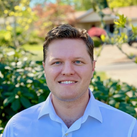
About Our Guests: Dr. Jacques Zaneveld is a pioneer in medical technology, with a background in human genetics and a passion for improving surgical training and patient care. As the founder of Lazarus 3D, he leads a team of scientists and engineers dedicated to advancing the field of surgical simulation. Dr. Zaneveld’s expertise and innovative spirit have led to the development of cutting-edge 3D printing technology that is transforming surgical practice.
Topics Covered: Throughout the episode, Jen and Dr. Zaneveld explore various aspects of Lazarus 3D’s technology and its applications in healthcare. They discuss the development process, regulatory considerations, and challenges in creating lifelike anatomical models. The conversation also touches upon the potential impact of this technology on surgical education, patient outcomes, and medical device innovation. Additionally, they highlight job opportunities at Lazarus 3D and encourage listeners to explore careers in this exciting field of medical technology.
Our Guest: Dr. Jacques Zaneveld: Pioneering Surgical Innovation for Improved Patient Care
Dr. Jacques Zaneveld is a visionary leader in the field of medical technology, leveraging his background in human genetics to pioneer advancements in surgical simulation. With a Ph.D. in human genetics, Dr. Zaneveld brings a unique perspective to the development of innovative solutions for surgical training and patient care. As the founder of Lazarus 3D, he has spearheaded the creation of cutting-edge 3D printing technology that allows for the fabrication of precise anatomical replicas from MRI and CT scan data. Driven by a passion for improving surgical outcomes, Dr. Zaneveld leads a team of scientists and engineers at Lazarus 3D, dedicated to pushing the boundaries of surgical practice through technology.
Dr. Zaneveld’s expertise extends beyond scientific research to encompass a deep understanding of the healthcare landscape and the challenges faced by surgeons and medical professionals. His commitment to bridging the gap between theory and practice is evident in Lazarus 3D’s mission to provide realistic surgical rehearsal experiences for practitioners at every stage of their careers. By empowering surgeons with the tools they need to practice on patient-specific models, Dr. Zaneveld aims to enhance surgical preparedness, improve procedural outcomes, and ultimately, elevate the standard of patient care.
Beyond his role as a leader in medical technology, Dr. Zaneveld is also a passionate advocate for innovation and collaboration within the healthcare industry. Through partnerships with medical institutions and device companies, he seeks to drive continuous improvement and innovation in surgical techniques and medical device development. Dr. Zaneveld’s vision for the future of healthcare is one where advanced technology plays a central role in optimizing patient care, improving surgical outcomes, and advancing medical education. With his pioneering spirit and dedication to excellence, Dr. Zaneveld continues to shape the landscape of surgical practice and medical innovation.


Episode Transcript
Jen Callahan: Hey, everybody, welcome back to another episode of The Skeleton Crew. I’m your host, Jen Callahan, and tonight I have a great guest with me. His name is doctor Jack Zaneveld. He’s joining me from a company called Lazarus 3D. And it’s in the healthcare field. We’re not exactly focusing on the radiology realm tonight, but it’s a super interesting product and one of a kind of its own currently on the market. I’m super interested to have this conversation with Jack and to share what’s going on with his company with you guys. Jack, thanks for being with me tonight.
Jacques Zaneveld: Oh, thank you so much for having me, Jen.
Jen Callahan: My pleasure. I’m going to ask you to give us a brief oversight, and then we’ll get more in depth with what Lazarus 3D does.
Jacques Zaneveld: Absolutely. Well, if you look at any area of human endeavor, people get better the more they practice, and especially the more they practice with exactly what they’re going to do. If you look at people driving race cars, they will practice a particular track many, many times to get the optimal time. And the same thing, if we take the best violin player in the world, they’ll rehearse a piece of music many, many times before giving a performance. Well, we’ve never before been able to rehearse a particular patient’s upcoming surgery, and that’s really what we’ve developed at Lazarus 3D, using a novel 3D printing technology that I developed originally in my kitchen in Houston, Texas, and is now being run and led by a team of scientists and engineers. We’re able to build exact replicas of a patient overnight built from that patient’s MRI and CT scan data. And these replicas actually cut. They bleed and they enable you to really do a full surgery on them the same way you would or you will on your patient potentially the next day.
Jen Callahan: Wow. That’s awesome. How did that idea even develop for you? I guess you were in practice because you went to med school as well, right?
Jacques Zaneveld: I got a PhD in human genetics, but a colleague of mine was considering an MD PhD, and I went to some medical training events, and I was a little surprised at the models that were available. You know, you had people learning how to do cystoscopies on water balloons and using pieces of fruits and vegetables like chicken thighs. It seemed like there was very little bridge between watching surgeries and starting to perform some aspects of it, to really taking the ball and running with it. One of the other things that is part of our mission at Lazarus is to help improve hands-on skills training for residents and medical students, as well as everyone in their career paths. And that was a product we were able to launch earlier because that didn’t require FDA clearance. And then just in 2023, we got our expanded FDA clearance, where now we can build models of individual patients for use in patient care for essentially every surgery performed in the US.
Jen Callahan: I could imagine that for patients who are undergoing surgeries, you know, for maybe tumors that are being removed or very complex surgeries, that this is extremely beneficial to the surgeons that are using it. Practice makes perfect in any sense, right? You know, they can already see they’re looking at a Cat scan or an MRI and they can see, you know, in a flat image. I mean, even if it’s a 3D image, it’s not hands on. I can only imagine how beneficial it is for surgeons. What’s the feedback that you get from them?
Jacques Zaneveld: Yeah, that’s exactly right. We are in principle working on a lot of very challenging procedures, sometimes with multiple practitioners coordinating care for a patient. And for example, one thing we hear sometimes from reconstructive surgeons is they don’t know what state the anatomy is going to be in when it’s their time for them to work on that patient. By doing a rehearsal, the entire team can coordinate and have a better idea of how exactly they’re going to tackle challenging problems. I think that some of the best markets for this include very complicated procedures like neuro oncology applications, a conjoined twin case previously, as well as things like live donor liver transplant where there’s a lot of very complicated anatomy and where the stakes are high.
Jen Callahan: I was on your website earlier looking, and I have to say that you showed different robotic surgeries using this, and I can only imagine me one doing it with your hands, you know, having a surgery, using your own hands, but then having to manipulate a machine. How much more careful you have to be doing that and being able to practice or like you guys say, rehearse makes it all that much better.
Jacques Zaneveld: Absolutely. And one thing that we didn’t fully anticipate initially, but using this technology can really help drive innovation in the surgical space. So obviously whenever society does a procedure on a patient, you have to be extremely careful and it has to be very, very well thought out. And supported in order to get approvals to do any sort of a clinical trial. But that’s at odds with goals of innovation in that, you know, it slows down the pace at which new ideas can be tested. Part of what we found through working with major medical device companies is that using these technologies can help them develop the next iteration of surgical tools, and with surgeons, can help them develop the next generation of surgical techniques. You can take a synthetic copy of a patient and say, well, what if we did treat it this other way? What are the results going to be? That’s very powerful, right?
Jen Callahan: You can almost experiment and see what the result would be, which is great. And you’re not doing it on the patient themselves where there’s risk.
Jacques Zaneveld: That’s exactly right. One of our early clients was Doctor Sandeep Keswani, and he has been investigating in utero treatments of Gastroschisis. We were able to engage with him to build models of these fetuses where the intestines had come out through a defect near the umbilical cord inside the womb, and work with him to develop techniques to treat that condition before the baby is born. And I’m really excited to share that. That’s now in human clinical trials, and it’s just really exciting to see that evolution as well as that partnership grow.
Jen Callahan: You guys take the CTS and or MRIs and you make these models that are, you know, mimic like a real person’s flesh. How did you come across something like how to develop something that mimics human qualities?
Jacques Zaneveld: I tried a lot of things, but first I was just making models in hard plastic. Some of the first applications of this technology were actually in the legal sector, because as a student with a 3D printer that my friends and I had built, all we could do was print in hard plastic using sort of the standard FDM technology. If anyone knows about that. I started building models before and after an injury, and then legal groups would use these to show how much damage was caused as a result of an accident or injury, as demonstrable evidence that could be presented to the jury. So that gave us an opportunity to get really good at analyzing data. A lot of that was spine data at the time, which is a lot more straightforward than soft tissue conditions, and also to get enough funding from that to be able to develop this soft tissue technology. And we tried all sorts of things, like I remember taking a cow liver literally and putting it in a blender, and then trying to see if we could come up with a glue that would hold the blended cow liver together so that we could turn it back into, you know, whatever shape we wanted. That did not work. But that’s just one example of one of the things we tried.
Jen Callahan: Oh my God, this is probably off the wall, but so are you cooking the cat liver or are you doing it wrong? I don’t know why I’m thinking of this.
Jacques Zaneveld: Uncooked, but yeah, we went to a butcher in Houston, and cow livers are really big. You can get a lot of material to work with. Not a lot of money.
Jen Callahan: I’m sure, though, that you had to develop many different styles of flash. I guess we’re calling it for different body parts that the models are being made for. Was that difficult to be able to come up with the different consistencies? I mean, because you finally figure out the one and now you have to figure out so many other ones.
Jacques Zaneveld: Absolutely. And that work is still ongoing. In particular, we recently received an award from the NIH. A grant to support research together with Texas Children’s and University of Oregon, which is allowing us to further investigate some of the material property questions surrounding our models. And so there are all sorts of tests that you can do where you take little samples and you pull them apart and you make indents on them using machines and all sorts of mechanical engineering that goes into trying to determine, well, what exactly does a bladder feel like, and then trying to recreate that in a synthetic model.
Jen Callahan: And then I’m sure I mean, if you’re looking at a bladder, you have to think, is it filled? Is it not filled? I’m thinking that it might feel different depending on if it has urine in it or not.
Jacques Zaneveld: Yeah. And that was actually one of the struggles with our FDA clearance because actual anatomy, if you build it realistically will move a bladder if it doesn’t have any fluid in it is in a collapsed state. The same thing happens with our models. So what we ended up doing in a test of some of the bladders that we were building for the FDA to show how accurate we could be, we suspended the whole thing in a solution that was the exact same density as the model, so that it would stay in a totally neutral configuration, and then we could analyze that and show that that to within the acceptable tolerances matched the. Patient data that we had received the submillimeter tolerances.
Jen Callahan: You’re doing all this to make it, you know, matches as best as possible, but then also to I mean, I loved watching the videos where, you know, the surgeons or whoever’s doing the rehearsing is cutting and there’s actually bleeding that’s going on, you know, blood and, you know, fluids that come out from the body that they’re doing, which is great. You guys obviously made it and put so much thought into it to make it as lifelike as possible.
Jacques Zaneveld: That’s really our goal. Perfect practice can lead to perfect performance, right? Our goal is to make that practice and that rehearsal experience as realistic as possible. And we’re really on the cutting edge of this. Most 3D printers are only able to do hard materials which are really primarily intended for visualization. This is one of the first technologies that allows for these hands-on experiences with soft tissues in a patient specific manner that’s intended for care, but this field is going to continue to grow in terms of the areas of medicine in which it’s used, the accuracy and realism of models, including fluid dynamics as time goes on. One remaining challenge, for example, is heart valves. Even being able to resolve the shape of a patient’s heart valves from scans can be very challenging, as can actually producing them in a model. So that’s something that we’re very excited to be pursuing additional research and development around together with partners across the country.
Jen Callahan: This episode is brought to you by X-ray Tech. Org, the Rad Tech Career Resource. If you’re considering a career in radiology, check out X-ray Tech. Org to get honest information on school’s degree options, career paths, and salaries. The heart valve thing I can speak from experience, just I work in a hybrid or room where they do a taper procedure, where they’re placing the valve up through the femoral artery, and they have measurements based off of the Cat scan that they believe. You know, we need to have the x-ray tube angled this way and then up towards the head this way so that they can visualize the valve properly. And a lot of times not a lot of times. But there’s good cases where they have to change the direction of where we’re looking at it. You know, they might have thought that we needed it to be towards the right, but we’re actually going towards the left and where they think they have to go up towards the head. We’re actually moving the x-ray machine to go down towards the feet to visualize it differently. I can speak to that from experience for sure.
Jacques Zaneveld: And that actually sounds like a step that could potentially be optimized through use of these types of technologies. Our models x-ray and we’ll show a difference from surrounding tissues. You could actually position and see that ahead of the patient actually being in the O.R. One thing that’s really clear from all the data is that time spent when it isn’t actually a surgery is so much cheaper than time spent when it is really a surgery. Any way that you can optimize efficiencies in the O.R. has the potential for savings, as well as the potential to bring additional clinical value.
Jen Callahan: Is the pressure model. Have you guys kind of dedicated it towards or honed in on one specific body part or body area that, you know, your kind of trying to be 100% in and then moving on from there.
Jacques Zaneveld: With our expanded FDA clearances, there are four primary areas that we’re investigating right now. Those include transplant medicine, in particular live donor transplant procedures as well as congenital defects, various heart models and various neuro models, especially brain oncology applications. Part of our goal, we just got those expanded clearances in 2023. Part of our goal for this year is to test all four of those markets and determine, you know, which areas we can have the most impact the fastest, and then hone and refine our strategy from that.
Jen Callahan: Wow. For the congenital and, you know, the twins, I mean, that I can’t even imagine how intricate that is. And are you using like Cat scans and MRIs, usually in tandem to come up with these models?
Jacques Zaneveld: Yes, it depends on the type of surgery, of course, but we’ll typically receive the entire radiological workup for the patient. And we’ll typically use the best scan for the structures that we need to resolve. So often we are using MRIs for some structures and CTS for others. And we’re essentially always using different individual data sets. Right, to get the venous phase. Or is this arterial phase to get better resolution on all of those structures, as.
Jen Callahan: I do want to ask, when I was looking through the website, it said, you know, I guess this was geared towards more like patients, you know, ask if you’re a candidate for surgeries. Are there certain cases that really wouldn’t be considered like best case candidates as opposed to others that are absolutely?
Jacques Zaneveld: Well, we defer to physician judgment, of course, on where and when this technology would be most effective. In my opinion, it’s most helpful when there are complex cases or when there are practitioners involved that are still on the learning curve for that new technique or technology. If something is very standard, very run of the mill, I would not encourage people to use this technology. It’s likely an unnecessary step, but any time there’s a challenge that we’re looking at, that’s where we would love to, you know, offer to help.
Jen Callahan: This is just like a blanket question. Just because, you know, insurance is involved in everything. Is this something that insurance would possibly cover or is it on the patient or on the doctor?
Jacques Zaneveld: There are category three CPT codes covering the use of 3D models in patient care. Those are tracking codes. They may or may not be covered by any individual insurance provider. And the way that those codes end up entering more common usage is through physicians typically repeatedly requesting reimbursement under those codes and escalating those requests. That’s really what’s going to determine the degree to which this becomes a standard thing to cover from insurance providers.
Jen Callahan: Okay, so the more the reimbursements requested, the more likelihood that insurance companies will continue to give the reimbursement or approve it.
Jacques Zaneveld: Correct? Yeah, that’s a signal that they take which says, hey, doctors really want this. We should probably pay attention. And then at that point they’ll come to the table and negotiate. We’re still in the fairly early stages. Well, we are receiving and have received reimbursements. It is still a very new technology and the typical default answer is no. That’s something that you have to overcome with any new technology. There is one special case which is transplant medicine, where we believe this falls under 1 to 1 coverage under Medicare.
Jen Callahan: When you say for transplant medicine, are you talking more so about the removal of the organ from the body or the placement of the organ into the body, or both?
Jacques Zaneveld: Primarily the removal of the organ from the live donor. And you think about it like a live donor liver transplant. That’s an extremely complex procedure where you’ve got, you know, your portal vein and hepatic vein and, you know, hepatic artery and your biliary system and potentially other complicating factors as well. In some of these cases, they’ll be removing, you know, 70% of the liver from a live donor. And it’s one of the most intense procedures that’s ever done on a patient that is fundamentally coming into the hospital fully healthy. There is a special mechanism under Medicare where costs associated with organ acquisition can be covered, and that would include surgical costs associated with care of the donor. Sounds very.
Jen Callahan: Intense.
Jacques Zaneveld: And it’s incredible that people do that. And donors are just such heroes. You know, everyone’s heart goes out to the people that are willing to give that much of themselves to another human being to help save a life.
Jen Callahan: So where are you guys looking ahead for the future? I mean, I know that you said you have these four groups that you’re, you know, specializing in and that you are moving forward with, but are you what’s next, maybe a surgical procedure that you’re hoping to nail down as well?
Jacques Zaneveld: I mean, our vision is really that this technology is available wherever and whenever surgeons need it and request it. That means we have to get good at building all high complexity surgeries. Now, the really nice thing about our technology is it’s highly adaptable. We can use the same processes, procedures, technology and equipment in order to build any organ in the body. And these capabilities are going to continue to grow over time, in some ways, almost more of a market acceptance and business strategy standpoint in that if someone comes to us and really needs something, we can already provide it in most areas of medicine.
Jen Callahan: You already have a prototype of it in some sort.
Jacques Zaneveld: Well, and that’s what 3D printing is, right? It’s rapid prototyping. What our technology allows us to do is within days take in any shape from any scan and build it. The turnaround times are insane. I think the shortest we did was for a kidney cancer application, and it was 16 hours between the physician handing me a CD with the data and us handing them the organ the next day.
Jen Callahan: Wow. What type of production company? I mean, not the company, but where is it produced? Like how is it? I don’t know, I’m just thinking of the whole manufacturing line and all of a sudden this, like, organ pops out. I just don’t know if that’s a more involved question than what we should get into, but I’m just interested nonetheless.
Jacques Zaneveld: Well, yeah, I’d love to share. There Are some proprietary details we can’t go into, but we have a 22,000 square foot manufacturing facility in Philomath, Oregon. A whole lot of our biomedical engineers are graduates of Oregon State University. And of course, we have an incredible team with people from all over the country as well. And we really have it streamlined through data intake. Data analysis will generate what’s called a digital twin. So that’s a computerized copy of the patient showing what we’re going to build. And this is also toggleable. You can turn off and on all the different layers and all the different tissue types. To really get a digital view of what to expect. We’ll typically ask the physician to approve that unless they sign for pre-approval to say yes. This is the thing that I want. And sometimes they’ll say, hey, can you also build, you know, the chest wall and include the ribs and exterior, right. If we’re building a heart or something. Then we’ll go ahead and design that. But once they approve it, it then enters the straight manufacturing and 3D printing steps. Once it’s there again very quickly. Typically, we’re able to deliver the product for our clients.
Jen Callahan: Wow, I think that’s great how you can put so much detail into it. Like you said, like building the chest wall, putting the ribs there, all the different vasculature. It sounds so involved and that the quickest that you ever did one was in 16 hours. I mean, it just shows how much you guys have honed this down and put so much time into it to almost perfect it.
Jacques Zaneveld: Yeah, our team is really dedicated, but I do still think we’re in the early stages. I mean, this has the potential to go a lot further and really impact so many lives. Till date this year, this technology has assisted in the care of about 35 cancers. Patients, which is good for a startup. But if we really want to impact overall outcomes, you know, nationwide, we’re going to have to be ready to scale that quite a bit. And I think there is potential. A number of studies have shown that the largest driver of outcomes, in fact, over 90% of the variability, for example, if we take kidney cancer, over 90% of the variability in margin rate. Whether or not you leave some of the cancer behind, complication rates and readmission rates are based on surgeon factors. So that tells us that the better prepared and the better a surgeon is able to execute that really can impact patient outcomes. Another parallel that’s interesting is in aviation, when the US government mandated, I think it’s around 2000 hours of simulation-based training prior to people becoming commercial pilots, there was about a 90% drop in fatal crashes over about a ten-year period. And that was also in part due to some other technologies that were implemented. But it’s really clear that that level of that ability for pilots to realistically rehearse in flight simulators had a huge impact on performance in the field, since this has never before been possible, our hope is to really explore where and when this technology can be most impactful on patient health.
Jen Callahan: Is this solely at this point offered in America, or I should say the United States, or have your kind of branched outward yet or just still in the early stages?
Jacques Zaneveld: We are primarily focused on the US. We sell skills training models which are not patient specific globally, and we do have a partner in Dubai and in the United Arab Emirates. Help us with sales of models there and fairly unique as a country in that they will largely just take an FDA clearance with minimal additional steps and allow you to sell medical devices there, which helped us do that quickly. As I started up. You know, you have to make very important decisions about how much and where you’re going to invest in order to allow access in other countries.
Jen Callahan: I’m sure that they know the process in America, you know, the due diligence that, you know, companies have to go through for FDA clearances, I’m sure very vigorous.
Jacques Zaneveld: Yes. You know, we’re lucky to have one of the more vigorous regulatory environments in the world as it should be. And our experience with the FDA has been fantastic. I have loved working with them. That isn’t something a lot of MedTech CEOs will tell you, but I’ve actually really enjoyed it. And part of that is that I think a lot of people have, you know, made rudimentary models, you know, out of plastic, using off the shelf 3D printers and what have you. And we really see this as being critical and it being so important that we get things right, that we are actually representing the patient data to the best of our capability and analyzing it correctly, and that we’re able to rigorously, scientifically demonstrate that that’s the attitude that our entire team has brought into this endeavor that we see as something that differentiates us from other ways. Some other groups use this technology, and I really appreciate that rigor, because I think if we’re going to progress this as a field, we need to be holding ourselves to the highest standard and even to higher standards than, you know, the FDA will impose. We need to get it right.
Jen Callahan: I think it would be really beneficial to our listeners here if we could share a video to show. Exactly. For those of you who might be listening on Apple or Spotify or, you know, just listening to audio, if you have the opportunity to go onto YouTube as well so that you can see what he’s going to share, or you can check out the website as well. It’s Lazarus 3d.com. But Jack’s going to go ahead and he’s going to share a video. Those who are watching on YouTube can see what these 3D printing models are like.
Jacques Zaneveld: Thank you so much Jen. And yeah. Well we can see in this video is a lump that looks quite realistic with blood dripping over the top and someone using a scalpel to remove this lesion. This is from a breast lesion. And part of what’s really impressive is how the tissue sort of string and holds together. One of the new technologies that we developed allows us to control the amount of bonding between different tissue layers. In reality our bodies aren’t a solid chunk, right? We have these planes that surgeons need to find between different tissues. So that’s something that you can see in the model as well as now a reconstruction where the physician is suturing closed that incision.
Jen Callahan: I’m sure that suturing is a huge thing, learning how to get the skin to, you know, line up with each other and you know, as much as you can practice, practice, practice. I don’t know if I mentioned this earlier, but I do like how you guys say rehearse instead of practice. It’s a good transition for words there. I do appreciate that because they think of someone like practicing on a human or practicing for your upcoming surgery. I feel like it’s not the best way to think. I like the word rehearse a lot.
Jacques Zaneveld: Well thank you, and if any of you are interested in suture training products, I’d recommend you check out the Suture Buddy. And we’re the proud manufacturer of products for them.
Jen Callahan: People who join your company, who exactly do you look for? Because, I mean, it seems like you would need a pretty good skill set to join this company. I definitely know that. I don’t think I’d be a candidate there, but who do you look for employee wise?
Jacques Zaneveld: Yeah, we’re a startup and we’re rapidly growing, so right now we’re posting a lot of openings. But those include in particular for the people on this podcast. We’re looking for people who have a lot of talent when it comes to analyzing MRI and CT scan data. People with expertise in segmentation and in creating these realistic and incredibly accurate models from that data, that is a skill set that we are looking for. I’d encourage you to reach out, call, email or check us out. Indeed, for our job postings, we’re also currently hiring for sales, for marketing, and for a number of different production positions including engineering, management, etc.
Jen Callahan: Okay, so people who would specialize in segmentation, would that be like a doctor, like a radiologist or a technologist or someone who specializes in QC for radiology?
Jacques Zaneveld: Potentially all of the above, but also medical illustrators are one example who are often generating 3D designs.
Jen Callahan: Well, everybody, you’re here in this first, just like I am about Lazarus 3D doing some monumental work out in the healthcare field, making lifelike replica bodies coming so that your surgery can be better than it ever could be, especially if it’s complicated. So, Jack, I really appreciate your time and sharing everything that your company is doing.
Jen Callahan: Absolutely. Thank you so much, Jen. I really appreciate it.
Jen Callahan: My pleasure everybody. Jack Zonneveld shared with us about Lazarus 3D. And if you’re in the market for a job, check out them for a job because it sounds really interesting. But everybody I’m Jen, this is Jack. Check us out on future and past episodes on Spotify, Apple, and also two on YouTube. We’ll see you guys next week.
Jen Callahan: You’ve been watching The Skeleton Crew, brought to you by X-ray Score.org. In the next episode, join us to explore the present and the future of the Rad Tech career and the field of radiology.
