Journey to Business and Mammography Success with Deborah Thames of MD Anderson Cancer Center
Episode Overview
Episode Topic: In this episode of Skeleton Crew – The Rad Tech Show, join Deborah Thames, a seasoned Mammography Technologist at MD Anderson Cancer Center. Discover the inspiration behind Deborah’s 25-year specialization in mammography and gain insights into the evolution of this critical field since 1996. Uncover pivotal moments at MD Anderson that highlight mammography’s crucial role in cancer detection and treatment planning. Explore Deborah’s transition to education, as she shares her motivations for teaching mammography courses and contributing to the training of future mammographers at UT School of Health Professions.
Lessons You’ll Learn: Explore the evolving landscape of mammography training with Deborah Thames. As she teaches the 40-hour initial mammography training, learn about the changing curriculum and the emphasis on emerging technologies. Gain valuable advice from Deborah, an expert in guiding accreditation processes, as she shares insights for aspiring mammographers and radiologists navigating accreditation requirements. Discover the integration of academic knowledge into practical mammography, as Deborah discusses the benefits derived from her recent Master’s degree.
About Our Guests: Meet Deborah Thames, the passionate advocate for breast health. Uncover how Deborah leverages her extensive knowledge to contribute to the fight against breast cancer. Explore the vital role she sees for technologists in raising awareness about breast health and regular screenings. Look into the future with Deborah as she anticipates advancements in mammography and shares how healthcare professionals, especially radiologists and aspiring mammographers, can stay informed..
Topics Covered: Embark on an inspiring exploration of Deborah Thames’ remarkable journey as a mammographer, educator, and breast health advocate. Uncover the invaluable experiences that have shaped her expertise. Discover how Deborah’s passion for breast health fuels her impactful contributions to the fight against breast cancer. Gain insights into the evolving landscape of mammography through her eyes. Express appreciation for Deborah’s unwavering dedication and be inspired by her commitment to advancing mammography. Radiologists and aspiring mammographers, this is your chance to glean wisdom from a true industry leader. Stay tuned for a powerful conclusion to an enlightening episode.
Our Guest: Deborah Thames, A Pioneer in Mammography Education and Advocacy
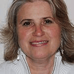
Deborah Thames is a seasoned radiologic technologist, specializing in mammography since 1996. With over two decades of experience, she has become a trailblazer in the field, dedicating 24 years to MD Anderson Cancer Center in Houston, Texas. Beyond her clinical role, Deborah has been a driving force in education and advocacy, making her mark as a lead technologist and a medical educator.
At MD Anderson, Deborah excels in screening and diagnostic mammography, stereotactic core biopsies, and digital mammography. Her proficiency extends to routine and fluoroscopic diagnostic procedures in diagnostic radiology. Deborah’s journey began with a radiologic technologist certification from the Stephen W. Brown School of Radiology in Augusta, Georgia, setting the stage for her impactful career.
As a medical educator at the UT School of Health Professions at MD Anderson, Deborah plays a vital role in shaping the next generation of radiologic technologists. She teaches the 40-hour initial mammography training curriculum and prepares students for the ARRT registry. Her commitment to education culminated in the completion of a Master of Science in Radiologic Sciences degree in August 2021.
Deborah’s influence transcends the classroom, as she travels nationwide to guide accreditation processes and MQSA inspections for MTMI. Her consulting role reflects a dedication to elevating mammography standards across the United States.
Deborah Thames stands out not only for her technical prowess but also as a passionate advocate for breast health. With 25 years of experience, she continues to contribute significantly to the fight against breast cancer. Through teaching, consulting, and her daily work, Deborah emphasizes the critical importance of early detection, education, and maintaining the highest standards in mammography.


Episode Transcript
Deborah Thames: Many mammograms are done incorrectly. That’s why facilities fail. Accreditation. They’re taught wrong. It’s not always their fault. It just saddens me because we’re here to save lives and find breast cancer at the earliest stage possible. So that, the mammogram is performed incorrectly. We’re not saving as many lives as we need to. Regulations state we have to do at least two views. Many times it is not done correctly where tissue is missed just from the lack of the technologists’ skills or lack of knowledge that’s given to these technologists. It just breaks my heart.
Jennifer Callahan: Welcome to the skeleton Crew. I’m your host, Jen Callahan, a technologist with 10+ years of experience. In each episode, we will explore the fast-paced, ever-changing suburbs, a completely crazy field of radiology. We will speak to technologists from all different modalities about their careers and education. The educators and leaders who are shaping the field today, and the business executives whose innovations are paving the future of radiology. This episode is brought to you by X-raytechnicianschools.com. If you’re considering a career in X-ray, visit X-raytechnicianschools.com to explore schools and to get honest information on career paths, salaries, and degree options. Hey, everybody, welcome back to another episode of The Skeleton Crew. I’m your host, Jen Callahan, and tonight I have a great guest with me, her name is Deborah Thames. She works at the MD Anderson Cancer Center as a mamotechnologist. Deborah, thanks so much for taking the time to be with me tonight.
Deborah Thames: Thank you for having me. So glad to be here.
Jennifer Callahan: Oh, perfect. We were just speaking to another Memotech a few weeks ago in education, which you also are in as well, so it’ll be a really good conversation to have. Alongside with the woman that I spoke with last week to see where we’re at in terms of your knowledge that you have, of what you’re teaching and the program that you’re in, and then also for me to know, how does your center work in terms of exams and the functionality of everything, and then being proactive in terms of screening for patients and stuff. So, really looking forward to our conversation here. Let’s get started. Can you give me a little bit of a background of yourself? You started being a technologist in like 1996, I think.
Deborah Thames: Yes, I went to X-ray school in Augusta, Georgia. I lived there for a little while. My husband had been transferred there, so I went to a hospital-centered X-ray program. And then I graduated in 1995 from the x-ray program. And my first job, we had to do everything CT, MRI, and Mammol. And I had never done mammography. I know in school that they showed a little bit of it, but there wasn’t MQSA back then because it was written in 1992 and implemented in 1994. So, When we were practicing in x-ray school, MQSA wasn’t even around then. I had to work on the mobile in Augusta, Georgia, and I really liked it a lot. I had really been introduced to Mammol, but I really liked it a lot, and that’s where I had got started with my interest in mammography. Then my husband had been transferred to Houston, Texas, because he was in the cell phone business. So, I tried to find a job here. I couldn’t find an X-ray job at all at the time. The X-ray techs were too many here in Houston, so I couldn’t really find a job. The only job I found was in mammography. I worked at this facility for a little bit then I transferred to MD Anderson Cancer Center. I just felt it was a better fit for my future career in mammography.
Jennifer Callahan: So when you first took that position in mammography, had you taken the boards for that or at that point, was it kind of like a cross-training situation where they didn’t require you to be registered in it?
Deborah Thames: There weren’t any rules back then to take any boards or anything, it was a volunteer. So, I did take the registry mammography registry in 1996. I hadn’t even really done mammography that much. I didn’t know a whole lot about it, I barely passed with a 78 score, but I did get my M in 1996. And then when I got to Houston, Texas, I needed a whole bunch of CEUs at one time. So my facility sent me to mammography school because you could get 40 hours very quickly in a week. So I did eventually go to mammography school here in Houston, Texas. But I had learned a lot in mammography school that I didn’t know. I had gained a lot of knowledge and better skills and tricks that it takes to do mammography.
Jennifer Callahan: Thinking about it, sometimes the hands-on approach is what is needed for information to actually sink in. So, maybe you actually already being in the field and then learning a little bit after learning the ‘Hows’ and ‘Whys’ behind something. Maybe, it was an easier click for you than sometimes going into the didactic work first, and then having to wait to do the clinical setting portion.
Deborah Thames: I agree with that, but that’s why I feel some of the laws in mammography need to be changed. They had changed some and I felt they changed backwards in a way because it’s sending someone out to drive a car when they hadn’t studied the handbook for driving. It’s almost backward sending technologists to learn mammography and sending some to patients first and then go to mammography school. And that’s okay. That used to be that way but it is now, I feel that I really want to go to Congress and change many laws with mammography through what I have learned about the initial training with mammography.
Jennifer Callahan: How does your center work at MD Anderson? I mean, that’s a really well-known name in the country for cancer research and treatment. I’m assuming that breast cancer is very prevalent right now, but you guys have a large patient load of patients that possibly are coming to see you from other parts of the country, or..
Deborah Thames: Oh, yes. We have a very large facility. We have a screening facility for cancer prevention programs where both men and women can come for skin and men for prostate and breast and gynae and colon, it’s a one-stop shop where they can get many things screened. So, we do many screening mammography exams down in Cancer Prevention Center, about 100 a day that we do. And then we do have a separate clinic that’s upstairs in a separate part of the hospital that we do diagnostic mammograms for patients who have breast problems, newly diagnosed staging, and also, women who come back for their annual exam that have a history of breast cancer. We also have an undiagnosed clinic where patients can get an appointment the very next day when they have breast problems because it’s important to get these types of patients in a hurry that have breast issues. We have special time slots for those that can be added on very quickly for a mammogram and a following ultrasound. We do about 80 patients upstairs in diagnostics in the clinic. Also, we have 4 mammography mobile vans that go out and serve the community. I have been on the mobile for about 22 years. We had to get our CDL license, our commercial driver’s license because the vans are over £28,000. So it requires us to have a special license to drive them. And two of us techs go out and go to clinics and serve the community. And many patients do not have insurance. So, we get grants through people who write for grants and then through special communities. Some other companies do raise money for patients who are underserved. Then we also serve businesswomen. We go to companies to where they don’t have to take a whole day off of vacation to get breast checks and like clinical breast exams and screening mammograms. So, we also go to companies around the Houston area and serve mammograms businesswomen who need screening mammograms. It just takes 15 minutes and sign them in and come down. And then they can go right back to work. So, we have a lot of services at our facility to reach out. And yes, we also have international cooperation with interpretation. We have legal interpretation for many different languages, for our patients who come internationally, so they can have services at our facility. Since, we are the number one cancer center in the world, we do get many patients from out of state and out of the city and international. All right.
Jennifer Callahan: So let’s transition then from you working in the clinical field to when did you find yourself wanting to move into education for mammo?
Deborah Thames: Well, at the time I had a boss who taught at a continuing education company and she said, we really need teachers. Why don’t you teach? I said, no, I am not a teacher. She begged me and begged me wouldn’t stop. So finally I said yes and my first class I taught was the eight hour fulfilled Digital Mammography requirement class and I liked it. It was a little scary at first, and I didn’t do well my first couple times. And I started practicing more and got more information into the classes and really loved it. So that’s how I got started. And I also had helped with positioning, hands on positioning courses she had done. So I had started doing that about 2002, and then I got into teaching about 2005 and 2006, and then she had quit and went on to another job. So I took over the 40 hour initial training for mammography and I’ve done that since about 2009, and then also at the University of Texas at MD Anderson. I wanted to help start a initial mammography training there because we have many great resources with plenty of patients. We have 16 mammography units, and we had the resources to create wonderful, high quality mammography texts that can go out and save lives. And that took a long time to do it. And finally, three years ago, it came to pass where we had started.
Deborah Thames: And as part of The Bachelor, they have a three year bachelor program and X-ray school. And the third year they make a choice of what they want to do, like CT or MRI or interventional. They have to choose their modality, specialty that third year. And so mammography and CT are combined. So it’s a great program. And we carry them all the way through their clinical requirements and education requirements for the registry also. And that’s that’s a lot to do. But it’s great. We have many students have gotten jobs. We just hired two new students. So you create these students that know your facility and what’s high quality. And so it’s great to hire these students. And we’re so blessed to be able to have them at our facility. When you know their work ethics, their education and what and they have learned the correct way, that’s one problem. In mammography, many mammograms are done incorrectly. That’s why facilities fail accreditation. Oh, okay. Because they’re taught wrong. It’s not always their fault. So many mammograms are done incorrectly in the United States. It just saddens me because we’re here to save lives and find breast cancer at the earliest stage possible so that the mammogram is performed incorrectly. We’re not saving as many lives as we need to.
Jennifer Callahan: Now you’re saying incorrectly, what exactly does that mean? Like the proper images aren’t done or part of the breast is missing? Or what do you mean it’s done incorrectly?
Deborah Thames: Correct. The part of the breast is missing. Regulation state. We have to do at least two views in screening mammography CC and Mlo views. And so both of those views are done. But they are done incorrectly because mQSA laws state we have to show 100% of the breast tissue. Well, they give us two views to do it in many times it is not done correctly where tissue is missed just from the lack of the technologists skills or lack of knowledge that’s given to these technologists. It just breaks my heart. It breaks my heart because they’re not trying to do bad mammography, but it’s just a lack of skills and education and knowledge about what needs to be on the image.
Jennifer Callahan: Now say, if a woman has a large breast or a part of the tissue was missed, and the tech did realize that you say that you have these two views to do it. You had a patient who was coming to have an abdomen x-ray, but they had a large belly. Sometimes you might have to do in what we call quadrant. You break the belly up into four pieces. You do the right, upper left, upper left, lower right, lower. You know what I mean? Is that the same theory that that you would use in mammography?
Deborah Thames: Yes. They call it mosaic imaging or tiling, where we do multiple images on a patient who has a large breast. So we know that we get the whole entire breast on there and all the views. So the minimum views we do on a normal breast average size patient is four views. Well sometimes we do 16 and 20 images to get 100% of that breast tissue on the image we get the pectoralis muscle. It’s very important. The pattern of the muscles correct. It needs to be wide so we can see some lymph nodes. Some breast cancers do happen in the muscle are attached to the pectoralis muscle. So we have to show this to the radiologist. So it’s very important to get the pectoralis muscle in the views on the. We try to get it on the view, but really only about 30 to 40% of the time we can get it on the view which is top to bottom.
Jennifer Callahan: And I’m sure that can be difficult. I mean, depending on, say, if a woman might be small chested or maybe the breast tissue is dense, I mean, being able to squeeze back towards where the muscle is, I can imagine that can be difficult where things can possibly be missed. Right?
Deborah Thames: Yeah, it could be difficult. But the law state you have to do it no matter what body habitus the patient has. The law doesn’t say, oh, you have a thin muscular patient, so you don’t have to do as good as mammogram. No, the law doesn’t say that. If a patient’s had a stroke, the law doesn’t say, oh, that’s okay. You don’t have to get a good mammogram. Now, you still have to get 100% of the tissue. So there’s supplemental views. There’s that we can do and there’s many additional views that we can do. And we just write it down. Why we did additional views or supplemental views to be able to accommodate this patient. Because it’s very important, no matter what physical disabilities they might have or even mental disabilities. We have done patients that have mental disabilities. And it doesn’t matter. We have they’re just as important to be able to get a high-quality mammogram. So we have to use skills and use multiple technologies. Sometimes there’s two and three technologies in helping with this one patient so we can accommodate that patient, do the highest quality imaging that we possibly can.
Jennifer Callahan: So when did you start teaching? At what point in your career?
Deborah Thames: In 2006, I had started teaching at one facility and then the person had quit. So I took over the mammography school, but I had written up to about 40 different classes with all mammography topics. And then I had changed companies about four years ago, and I helped with their mammography initial program also, and I also do for about 20 years I’ve helped with accreditation failures with the ACR and other accreditation bodies like the state of Texas and the state of Arkansas. Also, when facilities fail inspection, I can go in and help them correct their deficiencies and teach them how not to do something like that again, or teach them the rules, because many facilities don’t know the rules in mammography. It’s so strictly regulated and so many times also, we have to go in and do hands-on training with the accreditation process that’s required. And I’d love to go into other facilities because I learned from them too. I see their ways of doing things that could be different from our facility. So I get many new ideas from visiting all across the United States with teaching and going in and helping with the accreditation processes.
Jennifer Callahan: So the failure for accreditation is that for facilities like mammo screening facilities, or are you talking in terms of schools accreditation?
Deborah Thames: It’s mammography facilities. They must be accredited, certified and inspected to be able to perform mammography. Sometimes those processes, they fail and then they need help learning how to do mammography. Correct. And maybe some other things like. Quality control. We go in and help with quality control to help with the quality of their units. And there’s many different manufacturers, so we have to teach ourselves the machines and make sure when we go into these facilities, not only are we just teaching them about the patient and positioning, but we’re also teaching about the machine and how to get the best images out of their type of equipment that they have.
Jennifer Callahan: So once you go in and you do that, do you give them a certain amount of time before they’re re-examined on what they’re supposed to be up to par on?
Deborah Thames: They usually have 45 business days to redo everything. They have to re-send in images to different types of patients. One patient with dense breast tissue, and one patient with fatty tissue on every unit that they have. So they usually have 40 another additional 45 days to recorrect it and then send it all back into their accreditation body, whoever that is.
Jennifer Callahan: Since you started teaching in the early 2000, do you feel like the curriculum in the education centers that you’ve worked at? Do you feel like it’s evolved quite a bit?
Deborah Thames: Yes, it has evolved especially through different breast modalities. When I first started, I did a little bit of zero mammography, where it was done on paper and blue ink, and then it transformed into film screen with cassettes and chemicals to help cross. That was brought into the United States in the late 70s, into the 80s. And that process was wonderful until digital mammography came around in 2000, that had changed regulations with mammography, learning eight hours of digital mammography, and then came digital breast tomosynthesis in 2011. So the requirements changed to learn that modality, which is really good no matter what modality that you use, you must have eight hours of training in that breast modality to gain knowledge from that modality.
Jennifer Callahan: Would you say at this point, the digital breast tomosynthesis is pretty much what’s used throughout the United States?
Deborah Thames: Well, there’s over 25,000 mammography units in the United States, and only 11,000 are digital breast tomosynthesis. So less than half right now as of December 2023, are digital breast tomosynthesis. Hopefully more facilities will get it. It has been proven through clinical trials and all the scientific data that has been done on digital breast tomosynthesis to get approved by the FDA to perform digital breast tomosynthesis images on patients, that it does find more breast cancer using 2D and 3D in combination, and it has reduced recall rate. It has less biopsy fees that are being needed. And you can see the lesion as a whole. And there’s so many benefits to digital breast tomosynthesis. And I’m very surprised that the nation hasn’t kept up with newer type of breast imaging as far as facilities, but I know it can be expensive. I know this, and I think that’s one of the biggest problems. Why there aren’t more digital breast tomosynthesis exams being done in the United States. Do you think.
Jennifer Callahan: That it will come to a point where, say, for instance, like digital imaging in x-ray? I feel like by 2019 it was put out that anyone using X-ray equipment were required to have digital imaging or you were going to possibly lose your accreditation. Do you think that it might come to that with mammography, that maybe the ACR or whoever’s in charge of the accreditation will say you’re going to have until this year to upgrade your equipment?
Deborah Thames: Yeah, I think one day it will be like that. In fact, November 2023 was the first time that there no film screen units left in the United States.
Jennifer Callahan: Oh, wow.
Deborah Thames: Yeah. So they don’t come out and say you can’t use them. They’re obsolete. They just get phased out by patient demand. The patients have great knowledge through social media, the internet, and they can educate themselves. And they ask, do you have the latest technology? They ask it is pushed out by patient demand, which is good. I agree with the patient. Why not have the best if it’s available? And for a long time insurances wouldn’t pay. Just like when we went from film screen to digital insurances at first would not pay, then eventually regulations changed with insurance. Says that the states mandated that, yes, the state entrances are going to pay. So eventually then the regulations came out where they had to pay. Well, the same thing with digital breast tomosynthesis. At first insurances would not pay for it. Facilities purchases, very expensive, highly engineered pieces of equipment for mammography and they couldn’t get any paybacks on it. So they did offer the exams and had maybe the patient pay just a little bit extra upfront. And then eventually of course, now insurances and Medicare pay for it. So it does take a while for that to happen. But I think eventually they will say, yeah, if you’re going to perform mammography, you need to use the latest breast modality. But it could hurt patients, especially in like rural areas that do a low volume of patients. They might not have that availability to go get a screening mammogram so many times. That’s why they don’t do it because wonder if it’s one person or to be able to save their life screening them so many times. That’s why they don’t do it. But I think eventually they will mandate for you to get the best equipment out there.
Jennifer Callahan: So breast cancer is pretty prevalent, and I assume it’s because the promotion of breast screening has become the norm at this point. Do you feel like MD Anderson, since they’re like the leading in cancer research and treatment, do you feel like they do a good promotion for women and or men to be aware of their breast health, and to come in and get their screenings at the age that they’re supposed to?
Deborah Thames: Yes, they’re getting the word out to remind women to get screening mammograms. And annually there’s billboards around the city reminding women there’s websites teaching them about BSE or breast self-examination and about CVS, which are your clinical breast examinations and also education with classes. They do have some classes, especially through Cancer Prevention Center. There’s also clinical trials that women can sign up for that we let the public know. About one time we were doing a spit test. Wrigley had donated tasteless gum and sugarless gum, and we had the spit in this cup. And they were thinking, well, maybe there’s something in saliva that can indicate that you’re at a higher risk for developing breast cancer over a lifetime. And there’s other many clinical trials that one can sign up to help the future generation with breast cancer. Breast cancer awareness just puts the little click in a patient’s mind that says, yes, oh, I need a screening mammogram. I haven’t done it in a year or two and it really is needed annually. We know the number one risk factor developing breast cancer over a lifetime is gender. A woman has a 1 in 8 chance compared to a man who has 1 in 1000 chance. So gender is the number one risk factor and number two is age. And as we get older, our genes can’t repair themselves as well. So age is a risk factor. And those are very important risk factors and reminds them to get in annually. So you get a mammogram done in October of 2023. So you had a mammogram done. Well if you don’t come back annually, and if you wait two years and say you get breast cancer in January, well, if you wait another year and a half, that could develop to a later stage, invasive type of breast cancer instead of finding it early. So that’s why annual mammograms are so very important to find it at the earliest stage possible where it’s treatable.
Jennifer Callahan: What is it about the age 40 that why do they recommend for a woman to get her first mammogram?
Deborah Thames: Because that’s the decade of age where breast cancer risk jumps higher. With the second decade and third decade of life, the percentage is very low. And in the fourth decade of life, the percentage jumps high.
Jennifer Callahan: Do you think it has something to do with changing of hormones in the body at that point of life?
Deborah Thames: It can. That is a risk factor with a number of periods you’ve had over a lifetime, what age you started your period, what age you ended your period. Because it does have to do with hormones. If a patient is taking hormone therapy, that can bring some risk. And so it’s all about the education of that. But yeah, it’s very important with 40 and over that you get an annual.
Jennifer Callahan: Being a mama technologist, how do you feel that you or someone. To raise awareness for their patients in terms of breast health. Is it a reminder before they leave, like remember to schedule your appointment. Maybe you schedule it right away, or reminding possibly a woman to do a breast self-exam.
Deborah Thames: Yes, we remind the patients that you need to schedule for the next year. It’s very important that you come back every year and we also teach them. It’s important that you go to the same facility. We say if you can’t come to our facility, choose a facility that you can go to. That way the records are there, they have previous exams for comparisons and they’ll have the reports. Also, we do help to be an advocate to educate the patients about returning for their mammogram the next year. We do remind them, we do talk to them. And then they our patients go to the clinics. And the clinics also remind them so they get 2 or 3 reminders and then some facilities do send a reminder card. That’s a neat process. It can be costly through the postal system. Some will do it through like electronic health record as a reminder that, hey, it’s been a year, it’s time for your mammogram. They can have warnings through the electronic health records also, or what’s called like mychart, where they can look at their personal health records, even access their images, which is a nice plus for the patients to.
Jennifer Callahan: So going from that, how we were talking about the digital breast tomosynthesis. So that was a development in 2011 and it’s still working its way, trying to get into a good majority of facilities. Like you said, it’s not even in half of them. Do you see more technology advancing? I mean, technology’s always pretty much advancing. But do you have any knowledge of where or what might possibly be in the future for mammography imaging?
Deborah Thames: Yes. There is a new technology out called Contrast Enhanced Mammography. Our facility does it. Many facilities do it in the United States, where we inject patients with iodine contrast, and we have to do their images within six minutes before the contrast rushes out of their body. That is a great new technology. It has a high sensitivity and specificity rate for finding abnormal. Also, molecular breast imaging that has been around for a very long time, but it’s improved with spatial resolution where you see more detail and also reduction in dose from the radioisotopes. They first started out with gamma cameras, whole-body gamma cameras. Then they went to BSG, which is breast-specific gamma imaging which was a dedicated mammal machine also. Then they went to PEM positive emission mammography with lower dose and better detail. And now there’s MBI. Mbi has been done. I’m not quite sure exactly how long I know think we’ve done at our facility for about five years. One of the problems was the companies kept on bankrupt with that made these machines for molecular breast imaging, but other companies had taken over. So we have different diagnostic tools to help mammography and these other diagnostic tools. Yes, breast ultrasound is wonderful and breast MRI is wonderful. But these other newer breast modalities are also helping find breast cancer at earlier stages possible.
Jennifer Callahan: So would that type of molecular imaging be used in conjunction I guess with a med tech mammography?
Deborah Thames: Let’s do it. But the nuclear medicine technologist does help us with quality control of the machine. And we send our patients to get injections right through our nuclear med. Even though we could do it, our technologists were trained. It was just a time issue.
Jennifer Callahan: And in terms of the contrast-enhanced imaging. So you inject a patient with the contrast and then is the theory behind it that it’s going to go to the area of if there is some type of mass or something.
Deborah Thames: Yes ma’am. It does. It enhances on the subtraction of the computer. So it takes a high KVP image and a low KVP image, and then it subtracts it with the computer software. And it will show enhancement that possibly is a cancer. And it has the high sensitivity and specificity rate for finding breast cancers. And they do compare it to MRIs. It is a supplement of MRIs, especially for patients who can’t have MRIs due to maybe body habitus or claustrophobic or have metal implants in their body. So it’s a great alternative to MRI because it is less expensive, it’s faster to read for the radiologist, and there’s more time slots.
Jennifer Callahan: If you could real fear for people who might be listening that aren’t actually working in the field of radiology. Just give like maybe like a one minute synopsis of high KVP versus low KVP imaging for.
Deborah Thames: A lower KVP range for digital mammography could be they use 26, 27, 28, and then a high KVP for CM would be like 49 for that contrast. Also, it uses a different filter. It uses a copper filter. The filters use a mammography using molybdenum rhodium, silver and then it uses aluminum for Tomo, but this uses copper. Now some manufacturers even use titanium. It is a different filter, and so it does take a low-energy image and a high-energy in image. Because you want to go 33 is the perfect attenuation number for mammography. So they go below 33 and they go above 33. And then they subtract it. And that’s where you see enhancements. And we have seen many cancers that have not shown up in. The other modality, but it’s very interesting. I do teach a class on that, and I try to teach myself most things about it. We do it. I have done it at my facility. I don’t do it on a regular basis, but it’s amazing to me. I get shocked sometimes with the little tiny non-index cancers that have been missed on other breast modalities.
Jennifer Callahan: So is this just done through an IV then like say like you have an IV placed in your arm. You inject the contrast and then within I guess like a few seconds you have to start the imaging.
Deborah Thames: Yes, it is through an IV and we have power injectors just like they do in CT. We inject it with saline and the contrast. We finish with saline, and then we have to wait two minutes for it to circulate throughout the whole body. You have to tell the patient you’re going to feel like you’re going to urinate on yourself. You’re going to feel tingly. This is all normal that you have to tell them the symptoms that they’re going to be feeling. And after two minutes that is circulated, then you have six minutes to complete your imaging.
Jennifer Callahan: What would be considered high risk for was it someone who has already been diagnosed with it and is in recovery? I guess also, to someone who might have a family history of cancer, what other markers make you high risk?
Deborah Thames: Having dense breast tissue puts you at higher risk. Also, if you have mutation genes, there’s many mutation genes out there that can raise your risk of breast and or ovarian cancer. The more common ones are Brca1 and Brca2 can raise your risk. Also. Yes, having first-degree relatives like mother, sister or daughter is first degree relative, multiple relatives having breast cancer or ovarian cancer, and also having a biopsy with high-risk pathology such as RH, atypical lobular hyperplasia or atypical ductal hyperplasia, or else’s lobular carcinoma in situ. There are high-risk pathology that can raise your risk of developing breast cancer over a lifetime. There’s many other ones that can raise your risk. So we put these patients in a high risk, maybe short-term follow-up or alternating MRI and ultrasound and mammal. We do put them in a different category and keep a good eye on them. And sometimes they are offered endocrine therapy or molecular therapy like tamoxifen or remedies to help reduce the risk. So they do see counselors. We also have genetic counselors. If someone’s at a high risk, they get cancelled. And sometimes they do get blood tests to be able to see if they do have mutation genes that can raise their risk of developing breast cancer over a lifetime, then that counselor also will give them options bilateral prophylactic mastectomies or short-term surveillance or molecular or endocrine therapy. So there’s many options that are left up to the patient and their families on how they want to decide to make that decision.
Jennifer Callahan: Well, Deborah, thank you so much for sharing your wealth of knowledge with me and everyone else that’s listening with us tonight. Everybody, this is Deborah Thames joining me. She’s a professor at two different schools, and she works in the clinical realm at MD Anderson. I mean, you are a busy woman, and I’m very thankful for you taking your time to to meet with me tonight and share your information and your wealth of knowledge with me in the world. So thank you so much.
Deborah Thames: Well, thank you for having me. It was my pleasure.
Jennifer Callahan: All right, everybody, this is Deborah Thanes. I am Jen Callahan, your host here at the Skeleton Crew. We will check you out next week. All right. We’ll see you later, guys.
Jennifer Callahan: You’ve been listening to the Skeleton Crew, brought to you by x-raytechnicianschools.com. Join us on the next episode to explore the present and the future of the Rad Tech career and the field of radiology.
