Revolutionary Medical Imaging: Spectral X-Ray Advances by KA Imaging
Episode Overview
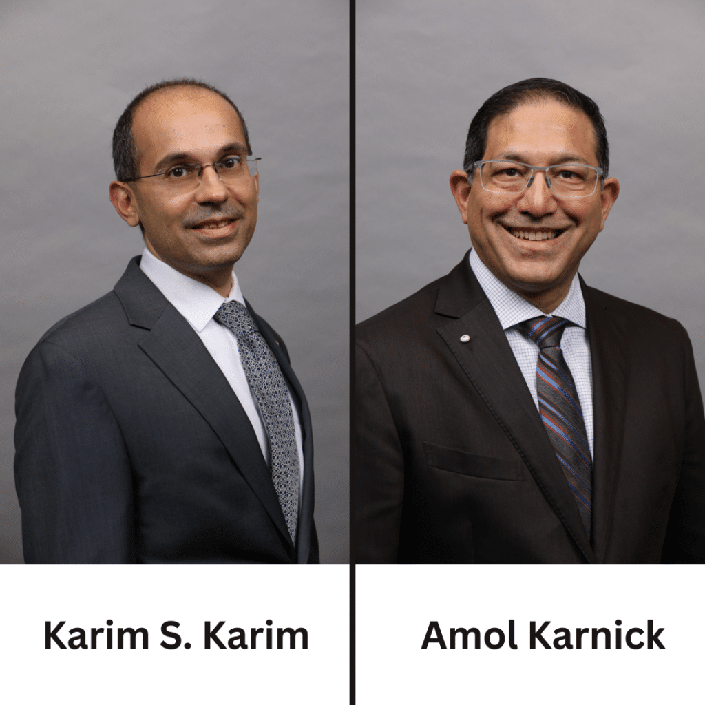
Episode Topic: In this episode of Skeleton Crew, host Jen Callahan engages in an insightful discussion with Karim S. Karim and Amol Karnick, co-founders of KA Imaging, a Canadian company at the forefront of innovative medical imaging technology. The conversation revolves around the groundbreaking technology developed by them, specifically their portable SpectralDR (spectral X-ray) detector. The episode delves into the transformative impact of spectral imaging on medical diagnostics, patient care, and beyond. Shedding light on the company’s journey, the technology’s capabilities, and its potential applications across various domains.
Lessons You’ll Learn: In this episode, you’ll learn about the exciting world of spectral X-ray imaging and how it’s different from traditional X-rays. How does this technology offer detailed material insights and shape data at the same time? The benefits of portable spectral X-ray detectors are that help boost radiologist confidence, lower radiation exposure, make workflows smoother, and even aid in catching diseases early. The guests also chat about the present situation of medical imaging, the difficulties it deals with, and the cool possibilities of combining AI with this technology.
About Our Guests: Karim S. Karim is a co-founder and CTO of KA Imaging and is a professor with extensive experience in entrepreneurship and medical imaging technology development. He has been a driving force behind the innovative advancements in spectral x-ray imaging. Amol Karnick is the CEO and co-founder of KA Imaging, he brings a deep expertise in engineering and medical imaging. His expertise lies in operations, sales, and engineering within the medical imaging sector.
Topics Covered: In this episode of Skeleton Crew, Jen Callahan interviews KA Imaging co-founders Karim S. Karim and Amol Karnick, and highlights their revolutionary work in medical imaging, focusing on the portable SpectralDR detector. They discuss spectral imaging’s transformative capabilities, including comprehensive material insights, improved diagnostics, and AI integration.
Our Guest: Karim S. Karim & Amol Karnick, Founders of KA Imaging Inc.
Amol Karnick, a seasoned executive in the medical device industry, boasts a diverse journey spanning startups like Ultrasonix, Sentinelle Medical, Ventripoint, and major players like GE Healthcare. With a background from the University of Waterloo and McGill, he has expertise in executive management, startups, mergers, and acquisitions. Notably skilled in sales and nurturing customer relations, he now serves as President and CEO of KA Imaging Inc., leveraging over two decades of experience to integrate sales, engineering, and operations, elevating the company’s industry presence.
Karim S. Karim is the innovative Chief Technology Officer of KA Imaging Inc. reshaping X-ray imaging since ’98. From Ignis Innovation to Ultrascan, and from ActivPixel to KA Imaging Inc., Karim’s journey echoes innovation. He’s harnessed $15M+ in grants, penned game-changing publications, and patented X-ray circuitry wizardry. His squad’s multi-layered detector is propelling dual-energy X-ray into a new era, upping clarity while dialing down radiation.
Delve into their universe at www.kaimaging.com where medical imaging evolution unfolds.


Follow KA Imaging on
Episode Transcript
[00:00:00] Karim S. Karim
X-ray radiation is also a spectrum and you have lots of little energies inside it. The challenge today is that those energies are not being utilized. In fact, when you look at an X-ray, you see a black-and-white image and it tells you something about the shape of the object that you’ve imaged. But actually, if you use the spectral information, not only can you get shape information, you can also get material information because the different energies interact with the different materials in the human body differently. And this allows you to identify calcium. It allows you to identify water iodine. So SpectralDR or spectral X-ray is the world’s first portable spectral X-ray device.
[00:00:42] Jen Callahan
Welcome to the Skeleton Crew. I’m your host, Jen Callahan, a technologist with ten-plus years experience. In each episode, we will explore the fast-paced, ever-changing suburbs. Completely crazy field of radiology. We will speak to technologists from all different modalities about their careers and education. The educators and leaders who are shaping the field today and the business executives whose innovations are paving the future of radiology. This episode is brought to you by X-raytechnicianschools.com. If you’re considering a career in X-ray, visit X-raytechnicianschools.com. To explore schools and to get honest information on career paths, salaries, and degree options.
[00:01:30] Jen Callahan
Hey everybody, welcome to another episode of The Skeleton Crew. Today I have two extraordinary gentlemen with me. They are joining me from Canada, which is awesome because I’m here in Philadelphia. Karim and I have Amol Karnick, who are from the company KA Imaging, and they have made some great advancements in the technology that they have developed and they’re going to take the time this evening to discuss that with me. So thank you, gentlemen, so much for taking time out of your lives to join me today to discuss this.
[00:01:58] Amol Karnick
Thanks, Jen. Appreciate being here. As you said, my name is Amol Karnick, president and CEO, one of the founders of KA Imaging. This is a quick background, As I say, I’m an engineer by training, but that’s about it. I did my undergrad engineering and master’s but ended up at GE Healthcare. So I’ve spent most of my career all in medical imaging, whether it be running operations, sales, engineering. So a lot of pretty deep background across the medical imaging businesses.
[00:02:25] Karim S. Karim
Now, thanks, Jen, for having us. My name is Karim. Also one of the co-founders for KA Imaging. I’m actually a professor by trade and got into entrepreneurship about 20 years ago. Three companies later, here is the lucky fourth and we can tell you lots about how we got started.
[00:02:40] Jen Callahan
All right. As I was saying in the introduction, they developed their company KA Imaging, and the detectors that we’ll be speaking of. Is it one of a kind currently on the market?
[00:02:50] Amol Karnick
Yeah, absolutely. It’s the world’s first SpectralDR or spectral X-ray detector.
[00:02:56] Jen Callahan
And do you want to just explain what the spectral means to our audience?
[00:03:00] Karim S. Karim
Absolutely. So SpectralDR, just like your eye can see three colors because white light has, you know, like a rainbow of colors inside it. X-ray radiation is also a spectrum and you have lots of little energies inside it. The challenge today is that those energies are not being utilized. In fact, when you look at an X-ray, you see a black-and-white image and it tells you something about the shape of the object that you’ve imaged. But actually, if you use the spectral information, not only can you get shape information, you can also get material information because the different energies interact with the different materials in the human body differently. And this allows you to identify calcium. It allows you to identify water iodine. So SpectralDR or spectral X-ray is the world’s first portable spectral X-ray device. And the advantages for the first time at the bedside, you can take this detector and you can get spectral information. You can find outlines and tubes. You can figure out pneumonias. You can figure out lesions like cancers, trauma, rib fractures, the whole lot.
[00:04:08] Jen Callahan
Is this also to something that is stationary within an exam room? You would mention bedside, but is there the technology is also in the exam room? So the same type of imaging you’re speaking of that you can do bedside, which is great because there’s many patients within a hospital that are sick, you know, and sometimes the exams that you’re performing could be not the best or somewhat compromise because of patient condition that this technology obviously aids.
[00:04:32] Karim S. Karim
Absolutely. The fact that you get material information. So the way it works is for every X-ray exposure, you get three images, you get the conventional image that you’re all used to. Then you get a calcium image which identifies all the bone and calcified arteries. Then you get a soft tissue image which lets you see the lungs and the lesions and the pneumonias and the pneumothorax and so on. So the beauty is this information allows you to make a decision that can reduce the need for a follow on CT. In a portable situation that can be very valuable. Think of ICU beds where patients are immobile. Think of trauma patients who cannot be moved. Think of long-term care homes and facilities where patients can’t be moved around, where transport is an issue. That’s where this type of technology makes a big impact.
The other place where this technology can also make a huge impact is in early detection or screening. We all know cancer is a problem. We all know heart disease is a problem. Those are the two biggest killers in the developed world. But early detection is kind of the one big thing that limits. For example, if you could find cancer at stage one instead of stage four, you have a huge advantage. If you can find heart disease at the age of 35, instead of waiting till you actually get struck down, that would be a huge advantage. Nothing today does early detection effectively, but with this technology, you can find lesions, indeterminate lung nodules, coronary calcium early with just an X-ray.
[00:06:04] Amol Karnick
And it really actually helps not just, you know, early diagnosis. It also helps the radiologist with their confidence levels. I’m sure you saw there was this report a few weeks ago that indicated some of the radiologist’s confidence level and diagnosis based on even standard chest X-ray was pretty low. And we’ve already shown through our own data and trials is that we’ve increased their confidence, plus enabled the intensivist, the emergency room doctors to be able to look at these X-rays with a higher confidence, make the decision at the time of need, rather than having to wait at times for report to get back to them.
[00:06:36] Jen Callahan
Right. They’re able to not say toggle, but they can toggle between the different images and looking at them from different viewpoints. Just coming off of my clinical background, basically, I feel like my understanding is you have your kVp and your mAs, and depending on which one is higher, you would use for different instances, one is more for bone, higher kVp or and then you have your other technique or soft tissue. So this instead of having to take multiple images with different techniques, you’re basically saying that the technology within this detector does that automatically.
[00:07:08] Karim S. Karim
Absolutely. And it goes one step further. It actually in the three images, it actually pulls out just the bone of the calcium. So you can just look at the bone without accompanying soft tissue. Right! Like when you look at a lateral chest X-ray, there’s so much soft tissue information. You miss a lot of things that you could be seeing, like a line could be easily hidden inside the soft tissue and you might miss it. With this technology, the soft tissue gets removed completely in the calcium image, And in the soft tissue image, the bone gets removed completely. So you get to see the material in all its glory without anything confounding sitting in there.
[00:07:43] Jen Callahan
It kind of similarly reminds me of an operating room setting, how the road mapping that doctors use, to blur out the bone so that they can just see the vessels.
[00:07:53] Amol Karnick
Yeah, we identify and separate the materials, making the visualization of what you need to see, not what you have to see to your point of what they do in the operating room.
[00:08:02] Jen Callahan
Right. So have you guys already implemented this type of detector?
[00:08:07] Amol Karnick
Yeah, you know, from a regulatory point of view so we can implement. We already have FDA 510(K). We do have actually Health Canada as well and also cleared in Europe and about eight other jurisdictions in the world. And we’ve got a few sites already installed in the US and they’re already actively using it and helping patients with better outcomes.
[00:08:24] Jen Callahan
I mean, it’s really, truly amazing. There’s patients who have who are intubated and you know, they’re getting like two, three, four X-rays sometimes a day looking to confirm the placement of, not just the ET tube, but other lines that that are placed as well, especially to if you think about nasogastric tubes that are going down into the stomach, patients who are kind of supine or recumbent, especially if they have a larger abdomen, you’re trying to penetrate through the abdomen.
[00:08:53] Amol Karnick
And we actually have a really good story on that one. We’re actually talking to one of the techs at one of the local hospitals in Toronto. And she was saying, you know, I was trying to find the NG(nasogastric) tube and I couldn’t with the regular X-ray detector and the X-ray system. So she went specifically to bring ours and she was able to easily see it at that point. You know, as you’re out there imaging yourself, you understand what you’re looking at. So this actually gives us, the techs, an ability to, you know, avoid retakes as well because they can see what they need to see. They know you’re going up there for an ICU patient. You know, they’re putting lines and tubes in placements. They want to make sure things are in the right place. The techs, the nurses, other people around the patient can actually get a better confidence in seeing what they can see and see those lines and tubes as well, and the PICC lines and the ports and making sure the tip is in the right place.
[00:09:35] Karim S. Karim
If we were to summarize the value that this type of technology brings into a hospital, it’s improvement in quality and risk reduction. Because if you can find problems, these patients are not going to be coming back because you will do the job right. For example, if you release somebody and they have a pneumonia, but you missed it, that pneumonia is going to get worse and they’ll be back in your hospital. If you’ve got somebody who’s got a small volume pneumothorax and you release them, well, they’ll be back within 30 days. And that also creates an exposure to malpractice risk, which this type of device would help reduce.
Then, of course, there’s the opportunities of incidental findings, which are really good for patients because you catch the disease early. It’s also good for the hospital because that revenue from that patient comes earlier. And then in terms of operating efficiencies, we know that staff is in high demand and they’re all burned out from Covid. So optimizing the use of their time. So you’re not doing unnecessary procedures is a huge deal even for the doctors. Right? Less time is needed to read and interpret the hard-to-read portable X-rays and you can make a good decision at the bedside without having to wait a long time. So in short, you’re improving both hospital and patient outcomes. You’re increasing revenue through incidental findings, but you also protect from a hospital perspective, from an administrative perspective, you protect revenue by reducing wasteful outflows, by doing unnecessary procedures, malpractice, so on and so forth. Right! Things that you want to avoid. So in a way, it’s a better detection in the form of an X-ray and one I think that’s very timely.
[00:11:07] Jen Callahan
And to touch on what you had said about reducing the amount of exams, having to be performed by the technologists, but also to helps from a radiologist standpoint because they’re reading less. If you’re submitting less exams or studies to be read, obviously it helps with their burnout as well.
[00:11:22] Amol Karnick
Absolutely. And like we said, even the intensivists who are making some of those decisions in the ICU. They will be able to make those decisions, taking some of that burden off the radiologists’ report immediately as well.
[00:11:33] Jen Callahan
So I want to kind of just switch real quick because as I was doing my homework on your guys’ website and everything, not only are you into medical human care, but you also to do work with veterinary as well.
[00:11:46] Amol Karnick
So I always like to highlight our vision. So when we say our vision is we do innovative X-ray everywhere. As you know, X-ray has been around 120 years, but the fundamentals really haven’t changed. You know, it’s the same X-ray Rodigan showed 120 years ago is essentially the same X-ray you see today. Yes, it’s gotten better in image quality and refinement and detail, but the fundamentals of an X-ray source and a film are pretty much the same. So we’ve improved that. So that’s why we say innovative. We’re trying to do things that are different, but then everywhere encompasses that one global health. We’re not just trying to do something in North America, trying to make sure we can deliver better health care globally, but also X-rays used in industrial imaging, in security, in veterinary medicine, so or nondestructive testing as well in terms of the other markets as well. So veterinary medicine is definitely an opportunity that we’ve been talking to some of the local vets, some equines, you know, so looking at the small and large animals, the equines are very interesting as well because they’re looking for the health of a horse before it runs. Is their bone spurs essentially or bone edema? They sometimes have to send those horses to CT.
[00:12:50] Amol Karnick
We can see some of these things a little easier as well. So we’re trying to do things, you know, in that portable setting. We’ve improved the imaging at the portable setting as well. So the point of care in terms of, let’s say a hospital terminology, but a point of care use, we’re improving the imaging at that point of use. And so that’s where the veterinary is like it as well. You know, some of them a lot of these vets also don’t have CTs. And we’ve shown that we’re showing imaging that we’re not exactly comparable to CT because we’re not 3D. But we have clearly shown that we’ve avoided other CTs. So if you can, for those vets to make a clinical decision on that animal, whether it be a dog, a cat, hamster or, rabbit, or whatever they have on the table for that day, and without having to refer that patient now to a CT somewhere because they weren’t able to diagnose it, they’ve actually kept that revenue as well and also kept that patient. They’ve kept that client happy in terms of being able to provide a diagnosis immediately on the spot with a high confidence as well.
[00:13:43] Jen Callahan
So you had touched on the veterinary and then also to using the X-ray. And so are you looking to incorporate into security-type areas?
[00:13:52] Amol Karnick
Actually we already have we made this as a public announcement. So I can say this. We did get some investment by In-Q Tel, who is the CIA investment arm. So that’s about as much as we can tell you. That’s a very interesting interaction with them. But they are using it for some security purposes. I love using this statement because this is a verbatim to what they told us. We loaned the detector to the government partner. They sent back to the In-Q-Tel team a simple email. It rocks! And so I love repeating that phrase because it was their words. And what they were able to do is because we show these different materials and identifying materials and separate the materials, we can highlight better what they’re looking for, whatever the threat may be that they’re looking for, they have a higher confidence against coming down to confidence. So when you look at our images, we give you the ability to see better. That’s by identifying different materials and showing them separately. And that’s where you can see better and that’s what they really like about it.
[00:14:46] Karim S. Karim
It’s also the same for things like aircraft parts. Aircraft wings are usually made of titanium composites. So with this type of detector, you can separate the titanium from the composite because composite is a low atomic number material much like water, actually even more like carbon. And then titanium is like calcium. It’s a little bit on the higher atomic number side. So you can actually split the two apart. You can do that for even buildings. If you wanted to separate concrete from steel, for example, trying to identify copper. So there’s actually a lot of applications where the multi-materials and the two materials have different atomic numbers. And this technology works really well there because as long as the two atomic numbers are low and high, you can kind of pull them apart and you can improve visualization, as I’m all said. And obviously, from identification of the material, you can also glean a lot of other insights.
[00:15:37] Amol Karnick
Yeah, it was interesting. I was having some conversations recently and bomb detection, you know, they’re looking at threats and packages and, you know, they’re trying to look for even a simple detonator or triggers, including an organic explosive aspect of it as well. So they’re looking at our technology doing well with this. We can identify the different parts and we know what the different parts are. And that really helps identify this is a bomb and how do we make we can make this as an automated bomb threat detection as well.
[00:16:01] Jen Callahan
When I was reading through both of your profiles, something that really caught my eye was about the fingerprint technology.
[00:16:08] Karim S. Karim
Yeah, years back when I was doing my PhD, this was probably about 20 years back. I developed a circuit technology for X-ray imaging. Now the funny thing is the medical companies never took it up. But years later, I got a call from an American company and they said, Oh, we heard about this technology. We’d love to use it for a different application. So what do you guys want to do? And. Well, it’s fingerprint scanning and said just fingerprints. And they go, Actually, no, we want the whole hand. And it was a security company. And so I worked with them for four years. They were based out of actually Amherst in New York near Buffalo, actually. And they took that technology and we worked together for four years and they actually made it work. And eventually, they ended up getting acquired. And behold, that technology ended up in cell phones. So every time you see an Android cell phone, if it’s ultrasonic biometric fingerprint reader that’s got that circuit technology developed early on.
[00:17:07] Jen Callahan
So you’re probably the second to third company that I’ve spoken to who are doing innovative technology like this. And it’s to me at least, mind-blowing the minds that are out there who can develop technology like this and be so creative because you truly are creative and your thought process. It’s amazing to me. I mean, I’m just like sitting here listening to you guys just so interested. So we’ve touched him a bunch of different stuff of what Spectral currently does, but let’s look forward to the future. And Kareem or Amal, would you like to share with us, Is there anything that you have on the horizon that you guys are hoping to work on or it might already be in the works?
[00:17:46] Amol Karnick
Absolutely. We definitely have multiple things in the works, and as an innovative company, we try to make sure we stay on top of what’s in the market, trying to make sure we’re delivering more, delivering more value, not just because we want to bring innovations, but we want to make sure that whatever we do make an impact, an impact in either outcomes for the patient or the clinicians to help them as well. And so today we’ve got a static detector. You know, it takes a single picture.
Our next version will be more like the fluoroscopy replacements or, you know, the dynamic versions where in real-time, you know, for if they’re doing a cardiac procedure, for example, they’re looking at the eye line. We could separate the eye down and highlight the iodine only, maybe suppress the ribs and look at the heart and the iodine together, suppress the ribs, or do other interventional procedures where they’re always using guidance. We can improve the guidance capabilities because we’re highlighting maybe the cars are coming in, we’re highlighting the bones or highlighting the structures that they’re looking for, or maybe the soft tissue they’re going after. As you probably know, with more and more minimally invasive techniques in terms of procedures that are happening, they need more guidance and they need more X-ray guidance or imaging guidance. And so what we’re trying to do is improve our imaging capabilities, especially in the OR and minimally invasive procedures to allow for better outcomes for the patients and for also making it easier for the surgeons to do their jobs.
[00:19:02] Jen Callahan
And it seems like with the technology that you guys have developed, sometimes with minimally invasive procedures, at times the X-ray dose could be higher. But because your technology can separate the two so that you would be planning on, you know, you said separate the heart, the iodine, but then you could possibly add the bone back on top of it if you want to.
[00:19:21] Karim S. Karim
You got it. You’re going to save on radiation. Even in a portable. We can save on radiation because our detector technology has actually three sensing plates inside each detector. So a conventional detector has just one plate. We’ve got three because we’ve got three. Our plates absorb more X-rays than any other plate in the market. So we give you a dose efficiency advantage even for a single portable x-ray. And now for fluoro, of course, the fact that you’re getting three images you can process, you can do all sorts of things. You don’t need to take two scans, you can take one scan and that’s a huge difference.
[00:19:57] Jen Callahan
So obviously, we’re talking about advantages to technologists and to doctors, but to our techs that are listening to us, besides the fact that like, it’s cool that I can separate these two and I’m going to decrease the amount of X-rays I might have to take. But are there any other advantages to us out there?
[00:20:16] Karim S. Karim
Absolutely. So you don’t have to change your clinical technique. You can use the same eyes that you’ve been used to. In fact, we even have a retrofit solution where you could take your existing x-ray source and just use our detector and software to get all the benefits of spectral. So we’ve got systems like a complete mobile system, but we’ve also got retrofit solutions where you just change the detector. That’s all you have to do.
[00:20:39] Amol Karnick
Like you say, Cream is highlighting this, the workflow side of it. It’s really important because, you know, we’ve talked a lot of techs and they don’t want to change workflow right now. And given the staffing shortages, the time and the constraints to learn something new is a challenge. So having the capabilities of not changing anything, we look and feel like anybody else’s X-ray system or even detector for that matter. So we don’t change workflow, we don’t change technique, but we give you better outcomes. So this is where, you know, we were talking about shortages. You know, think about the days when you’re doing the portables. You may have to go back and do a retake. We’ve already shown that we’ve avoided follow on imaging, so it’s less work and workload as well for the technologists. So that’s where we’re helping as well.
[00:21:16] Jen Callahan
Right. And just to catch on what you guys are saying, for those who might not be technologists, don’t know exactly what we’re understanding what we’re saying about technique. It’s like taking a picture and adjusting yours. To produce a different type of picture. But from technology, from our standpoint. I mean, anybody that has worked in different health systems probably knows that you might be going from health system A to health system B, and the one person has Siemens and the other one has Carestream and they run on two different numbers or exposure indexes. Your might be low over here, but then you’re going to have to adjust it to be higher over there. So basically these guys are saying that there will be no change in that it’s your company or if the house is dumb that you work for brings us in, which for me again, is mind-blowing and phenomenal.
[00:22:01] Amol Karnick
But a shameless plug if your people out there want to try it, we’re happy to support that as well.
[00:22:05] Karim S. Karim
Yeah, we can tell you if you use it in your ICU, you’re probably, and depending on how many patients you have, you’re probably going to save anything from 3,000 to $10,000 a week on just excess procedure costs that you would have paid and not gotten reimbursed for just by using this technology. There’s actually a really good financial incentive to try it.
[00:22:25] Jen Callahan
That’s great. And then also to financial incentive in terms of staffing, staffing being low. And at this point, people are burnt out and kind of like slacking off the field. You don’t have to worry about having, say, like four people on portables because you’re not going out hopefully, and having to perform as many exams that you could possibly, you know, have three technologists on instead of the four.
[00:22:49] Karim S. Karim
Absolutely. There’s a massive improvement on staff efficiency. As we go forward into the new year, we’ll be bringing out some AI solutions that will build on top of what we already have. So with the additional data channels, the AI is going to be significantly more accurate than any of the AI available in the field today.
[00:23:08] Jen Callahan
I’m happy that you brought that up. The three of us were talking about behind the scenes about the AI. Do you guys want to touch on a little bit more where you think you might be going or we’re going to save that for a future conversation?
[00:23:18] Amol Karnick
We can high level give you a quick overview because I think like as Karim said, you know, data, as most people know, data is the most important part for AI and the data that you get in really drives on what your outcomes are going to be. And every other AI in the world has all been trained on one X-ray image to train it. Fundamentally, we actually have six images. So for that standard X-ray, we get because of we have these three layers and the way we do the processing, we actually have six data points for every chest X-ray. We’ll be training with six data points rather than one. And plus we can identify the materials which other X-ray can’t. So we know our outcomes will be significantly better than anybody else in the market using our hardware.
[00:23:59] Jen Callahan
Are you thinking that it might possibly just go hand in hand with the way that the image is produced or that something that would come along towards where the radiologist who is reading it to use it as a companion?
[00:24:11] Amol Karnick
I think it’s both because we’re also seeing, as you said, you know, a lot of bedside diagnosis is happening or point of care is required and understanding that image at the point of care. So it enables the other physicians who are looking at the images, the technologists are looking at the images, you know, understanding is this normal, not normal. So do I need to take maybe another view? Do I need to, you know, call the radiologist right away to get them involved? Because there’s something here that’s really not normal. And this is an ICU patient. We need to make some decisions sooner so we help drive better decision-making. So what we’re looking at as well.
[00:24:42] Jen Callahan
This has already been put into play in different clinical settings. I assume you said up in Canada and a few already in the US. Have you met any resistance from, say, any doctors or technologists?
[00:24:54] Amol Karnick
I think our biggest challenge is to overcome the negative impact that technology you know, dual-energy X-ray used to be out there. So we’re fundamentally based on that technique. They heard about the negatives, about dual energy had motion artifact and double the X-ray dose. You couldn’t do lateral views. So they hear what we do like, Oh, it’s this dual energy. Well, no, that’s what we call it spectral. It’s not dual energy. So we have to overcome some of the negative connotations that dual-energy already has out there, even though it’s well clinically proven that it significantly improves outcomes. It’s just one of the challenges. I think that’s probably our biggest negative is just overcoming, you know, preconceived notions on what they thought of what this technique meant. So this is the reasons that we actually call it spectral X-ray or spectral doctor, similar to spectral CT, which seems to help overcome some of those earlier barriers that we had. Other barriers right now, which is being across the industry, is just, as you mentioned earlier, staff burnout and, you know, just availability of staff to look at something new. They’re just tired and under-resourced.
[00:25:56] Jen Callahan
The negative connotation. Now, it’s interesting because people have heard something for so long over and over again, even sometimes people with the thought of x-ray, but there’s been so much development that has occurred over the past 120 years. If you had said that the dose that’s received is minimal compared to what you might actually holding your maybe cell phone up to your ear constantly, maybe you’re receiving more radiation from that than just going to get a routine chest X-ray.
[00:26:25] Karim S. Karim
I mean it’s a different type of radiation but if you were to compare X-ray to a flight, if you’re taking a flight from the East Coast to the West Coast, that’s probably more dose than what you would get in a chest X-ray. So people sometimes forget that, right, because they’re taking flights all the time. But it’s actually got more cosmic radiation that could be absorbed in the body and cause damage to cells that the X-ray, you’re getting even less than that.
[00:26:50] Amol Karnick
All the flights that we got to take these days.
[00:26:53] Jen Callahan
Well, gentlemen, I want to thank you so much for taking the time with me today. I really enjoyed our conversation and I hope that we speak again in the future because I would love to hear more about what you guys have going on in the development.
[00:27:05] Amol Karnick
Thank you for your time. We appreciate being invited and have the opportunity to speak with you and your audience. And as you mentioned, if your audience members are interested, feel free to reach out.
[00:27:14] Karim S. Karim
Thanks very much for having us.
[00:27:15] Jen Callahan
My pleasure. So, everybody, I have with me, Kareem S. Kareem and Amol Karnick, and they are the CTO and the CEO of KA Imaging. All right, guys, hope to talk to you soon.
[00:27:32] Jen Callahan
You’ve been listening to the skeleton crew brought to you by X-raytechnicianschools.com. Join us on the next episode to explore the present and the future of the rad tech career and the field of radiology.
